[English] 日本語
Yorodumi
- EMDB-22528: Succinate: quinone oxidoreductase SQR from E.coli K12 -

+
Open data


ID or keywords:

Loading...

-
Basic information

Entry
Database: EMDB / ID: EMD-22528
TitleSuccinate: quinone oxidoreductase SQR from E.coli K12
Map data
Sample
  • Complex: Succinate: quinone oxidoreductase SQR from E.coli K12
    • Protein or peptide: x 4 types
  • Ligand: x 8 types
KeywordsComplex / Succinate: quinone oxidoreductase / SdhA / sdhB / SdhC / SdhD / ELECTRON TRANSPORT / ELECTRON TRANSPORT-OXIDOREDUCTASE complex
Function / homology
Function and homology information


plasma membrane succinate dehydrogenase complex / succinate dehydrogenase complex / succinate dehydrogenase activity / succinate dehydrogenase (quinone) activity / : / succinate dehydrogenase / cytochrome complex assembly / aerobic electron transport chain / anaerobic respiration / ubiquinone binding ...plasma membrane succinate dehydrogenase complex / succinate dehydrogenase complex / succinate dehydrogenase activity / succinate dehydrogenase (quinone) activity / : / succinate dehydrogenase / cytochrome complex assembly / aerobic electron transport chain / anaerobic respiration / ubiquinone binding / 3 iron, 4 sulfur cluster binding / iron-sulfur cluster binding / aerobic respiration / tricarboxylic acid cycle / respiratory electron transport chain / 2 iron, 2 sulfur cluster binding / flavin adenine dinucleotide binding / 4 iron, 4 sulfur cluster binding / electron transfer activity / membrane => GO:0016020 / heme binding / membrane / metal ion binding / plasma membrane
Similarity search - Function
Succinate dehydrogenase, hydrophobic membrane anchor / Succinate dehydrogenase, cytochrome b556 subunit / Succinate dehydrogenase, cytochrome b subunit, conserved site / Succinate dehydrogenase cytochrome b subunit signature 1. / Succinate dehydrogenase cytochrome b subunit signature 2. / Succinate dehydrogenase, flavoprotein subunit / Succinate dehydrogenase/fumarate reductase type B, transmembrane subunit / Succinate dehydrogenase/Fumarate reductase transmembrane subunit / Fumarate reductase/succinate dehydrogenase, transmembrane subunit / Succinate dehydrogenase/fumarate reductase, flavoprotein subunit ...Succinate dehydrogenase, hydrophobic membrane anchor / Succinate dehydrogenase, cytochrome b556 subunit / Succinate dehydrogenase, cytochrome b subunit, conserved site / Succinate dehydrogenase cytochrome b subunit signature 1. / Succinate dehydrogenase cytochrome b subunit signature 2. / Succinate dehydrogenase, flavoprotein subunit / Succinate dehydrogenase/fumarate reductase type B, transmembrane subunit / Succinate dehydrogenase/Fumarate reductase transmembrane subunit / Fumarate reductase/succinate dehydrogenase, transmembrane subunit / Succinate dehydrogenase/fumarate reductase, flavoprotein subunit / Fumarate reductase/succinate dehydrogenase, FAD-binding site / FAD-dependent oxidoreductase SdhA/FrdA/AprA / Fumarate reductase / succinate dehydrogenase FAD-binding site. / 4Fe-4S dicluster domain / Succinate dehydrogenase/fumarate reductase iron-sulphur protein / Succinate dehydogenase/fumarate reductase N-terminal / 2Fe-2S iron-sulfur cluster binding domain / Fumarate reductase/succinate dehydrogenase flavoprotein-like, C-terminal / Fumarate reductase flavoprotein C-term / Fumarate reductase/succinate dehydrogenase flavoprotein-like, C-terminal domain superfamily / Succinate dehydrogenase/fumarate reductase flavoprotein, catalytic domain superfamily / FAD-dependent oxidoreductase 2, FAD binding domain / Alpha-helical ferredoxin / FAD binding domain / Beta-grasp domain superfamily / 2Fe-2S ferredoxin-type iron-sulfur binding domain profile. / 2Fe-2S ferredoxin-type iron-sulfur binding domain / 2Fe-2S ferredoxin-like superfamily / 4Fe-4S ferredoxin, iron-sulphur binding, conserved site / 4Fe-4S ferredoxin-type iron-sulfur binding region signature. / 4Fe-4S ferredoxin-type iron-sulfur binding domain profile. / 4Fe-4S ferredoxin-type, iron-sulphur binding domain / FAD/NAD(P)-binding domain superfamily
Similarity search - Domain/homology
Succinate dehydrogenase hydrophobic membrane anchor subunit / Succinate dehydrogenase iron-sulfur subunit / Succinate dehydrogenase flavoprotein subunit / Succinate dehydrogenase cytochrome b556 subunit
Similarity search - Component
Biological speciesEscherichia coli (E. coli)
Methodsingle particle reconstruction / cryo EM / Resolution: 2.5 Å
AuthorsLyu M / Su C-C
Funding support United States, 1 items
OrganizationGrant numberCountry
National Institutes of Health/National Institute Of Allergy and Infectious Diseases (NIH/NIAID) United States
CitationJournal: Nat Methods / Year: 2021
Title: A 'Build and Retrieve' methodology to simultaneously solve cryo-EM structures of membrane proteins.
Authors: Chih-Chia Su / Meinan Lyu / Christopher E Morgan / Jani Reddy Bolla / Carol V Robinson / Edward W Yu /
Abstract: Single-particle cryo-electron microscopy (cryo-EM) has become a powerful technique in the field of structural biology. However, the inability to reliably produce pure, homogeneous membrane protein ...Single-particle cryo-electron microscopy (cryo-EM) has become a powerful technique in the field of structural biology. However, the inability to reliably produce pure, homogeneous membrane protein samples hampers the progress of their structural determination. Here, we develop a bottom-up iterative method, Build and Retrieve (BaR), that enables the identification and determination of cryo-EM structures of a variety of inner and outer membrane proteins, including membrane protein complexes of different sizes and dimensions, from a heterogeneous, impure protein sample. We also use the BaR methodology to elucidate structural information from Escherichia coli K12 crude membrane and raw lysate. The findings demonstrate that it is possible to solve high-resolution structures of a number of relatively small (<100 kDa) and less abundant (<10%) unidentified membrane proteins within a single, heterogeneous sample. Importantly, these results highlight the potential of cryo-EM for systems structural proteomics.
History
DepositionSep 1, 2020-
Header (metadata) releaseJan 20, 2021-
Map releaseJan 20, 2021-
UpdateMar 6, 2024-
Current statusMar 6, 2024Processing site: RCSB / Status: Released

-
Structure visualization

Movie
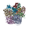
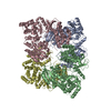
  • Surface view with section colored by density value
  • Surface level: 0.25
  • Imaged by UCSF Chimera
  • Download
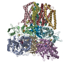
  • Surface view colored by cylindrical radius
  • Surface level: 0.25
  • Imaged by UCSF Chimera
  • Download
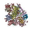
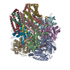
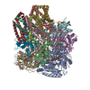
  • Surface view with fitted model
  • Atomic models: PDB-7jz2
  • Surface level: 0.25
  • Imaged by UCSF Chimera
  • Download
Movie viewer
Structure viewerEM map:
SurfViewMolmilJmol/JSmol
Supplemental images

Downloads & links

-
Map

FileDownload / File: emd_22528.map.gz / Format: CCP4 / Size: 10.2 MB / Type: IMAGE STORED AS FLOATING POINT NUMBER (4 BYTES)
Voxel sizeX=Y=Z: 1.08 Å
Density
Contour LevelBy AUTHOR: 0.25 / Movie #1: 0.25
Minimum - Maximum-1.543639 - 2.665797
Average (Standard dev.)-0.000000000000513 (±0.17199413)
SymmetrySpace group: 1
Details

EMDB XML:

Map geometry
Axis orderZYX
Origin109113104
Dimensions140137140
Spacing140140137
CellA: 151.20001 Å / B: 151.20001 Å / C: 147.96 Å
α=β=γ: 90.0 °

CCP4 map header:

modeImage stored as Reals
Å/pix. X/Y/Z1.081.081.08
M x/y/z140140137
origin x/y/z0.0000.0000.000
length x/y/z151.200151.200147.960
α/β/γ90.00090.00090.000
start NX/NY/NZ104109113
NX/NY/NZ140140137
MAP C/R/S321
start NC/NR/NS113109104
NC/NR/NS137140140
D min/max/mean-1.5442.666-0.000

-
Supplemental data

-
Additional map: #1

Fileemd_22528_additional_1.map
Projections & Slices
AxesZYX

Projections

Slices (1/2)
Density Histograms

-
Sample components

+
Entire : Succinate: quinone oxidoreductase SQR from E.coli K12

EntireName: Succinate: quinone oxidoreductase SQR from E.coli K12
Components
  • Complex: Succinate: quinone oxidoreductase SQR from E.coli K12
    • Protein or peptide: Succinate dehydrogenase flavoprotein subunit
    • Protein or peptide: Succinate dehydrogenase iron-sulfur subunit
    • Protein or peptide: Succinate dehydrogenase cytochrome b556 subunit
    • Protein or peptide: Succinate dehydrogenase hydrophobic membrane anchor subunit
  • Ligand: FLAVIN-ADENINE DINUCLEOTIDEFlavin adenine dinucleotide
  • Ligand: SODIUM IONSodium
  • Ligand: FE2/S2 (INORGANIC) CLUSTER
  • Ligand: IRON/SULFUR CLUSTERIron–sulfur cluster
  • Ligand: FE3-S4 CLUSTER
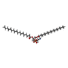
  • Ligand: 1,2-Distearoyl-sn-glycerophosphoethanolamine
  • Ligand: PROTOPORPHYRIN IX CONTAINING FE
  • Ligand: UBIQUINONE-2Coenzyme Q10

+
Supramolecule #1: Succinate: quinone oxidoreductase SQR from E.coli K12

SupramoleculeName: Succinate: quinone oxidoreductase SQR from E.coli K12 / type: complex / ID: 1 / Parent: 0 / Macromolecule list: #1-#4
Source (natural)Organism: Escherichia coli (E. coli) / Strain: K-12

+
Macromolecule #1: Succinate dehydrogenase flavoprotein subunit

MacromoleculeName: Succinate dehydrogenase flavoprotein subunit / type: protein_or_peptide / ID: 1 / Number of copies: 3 / Enantiomer: LEVO / EC number: succinate dehydrogenase
Source (natural)Organism: Escherichia coli (E. coli) / Strain: K12
Molecular weightTheoretical: 64.502766 KDa
SequenceString: MKLPVREFDA VVIGAGGAGM RAALQISQSG QTCALLSKVF PTRSHTVSAQ GGITVALGNT HEDNWEWHMY DTVKGSDYIG DQDAIEYMC KTGPEAILEL EHMGLPFSRL DDGRIYQRPF GGQSKNFGGE QAARTAAAAD RTGHALLHTL YQQNLKNHTT I FSEWYALD ...String:
MKLPVREFDA VVIGAGGAGM RAALQISQSG QTCALLSKVF PTRSHTVSAQ GGITVALGNT HEDNWEWHMY DTVKGSDYIG DQDAIEYMC KTGPEAILEL EHMGLPFSRL DDGRIYQRPF GGQSKNFGGE QAARTAAAAD RTGHALLHTL YQQNLKNHTT I FSEWYALD LVKNQDGAVV GCTALCIETG EVVYFKARAT VLATGGAGRI YQSTTNAHIN TGDGVGMAIR AGVPVQDMEM WQ FHPTGIA GAGVLVTEGC RGEGGYLLNK HGERFMERYA PNAKDLAGRD VVARSIMIEI REGRGCDGPW GPHAKLKLDH LGK EVLESR LPGILELSRT FAHVDPVKEP IPVIPTCHYM MGGIPTKVTG QALTVNEKGE DVVVPGLFAV GEIACVSVHG ANRL GGNSL LDLVVFGRAA GLHLQESIAE QGALRDASES DVEASLDRLN RWNNNRNGED PVAIRKALQE CMQHNFSVFR EGDAM AKGL EQLKVIRERL KNARLDDTSS EFNTQRVECL ELDNLMETAY ATAVSANFRT ESRGAHSRFD FPDRDDENWL CHSLYL PES ESMTRRSVNM EPKLRPAFPP KIRTY

UniProtKB: Succinate dehydrogenase flavoprotein subunit

+
Macromolecule #2: Succinate dehydrogenase iron-sulfur subunit

MacromoleculeName: Succinate dehydrogenase iron-sulfur subunit / type: protein_or_peptide / ID: 2 / Number of copies: 3 / Enantiomer: LEVO / EC number: succinate dehydrogenase
Source (natural)Organism: Escherichia coli (E. coli) / Strain: K12
Molecular weightTheoretical: 26.800912 KDa
SequenceString: MRLEFSIYRY NPDVDDAPRM QDYTLEADEG RDMMLLDALI QLKEKDPSLS FRRSCREGVC GSDGLNMNGK NGLACITPIS ALNQPGKKI VIRPLPGLPV IRDLVVDMGQ FYAQYEKIKP YLLNNGQNPP AREHLQMPEQ REKLDGLYEC ILCACCSTSC P SFWWNPDK ...String:
MRLEFSIYRY NPDVDDAPRM QDYTLEADEG RDMMLLDALI QLKEKDPSLS FRRSCREGVC GSDGLNMNGK NGLACITPIS ALNQPGKKI VIRPLPGLPV IRDLVVDMGQ FYAQYEKIKP YLLNNGQNPP AREHLQMPEQ REKLDGLYEC ILCACCSTSC P SFWWNPDK FIGPAGLLAA YRFLIDSRDT ETDSRLDGLS DAFSVFRCHS IMNCVSVCPK GLNPTRAIGH IKSMLLQRNA

UniProtKB: Succinate dehydrogenase iron-sulfur subunit

+
Macromolecule #3: Succinate dehydrogenase cytochrome b556 subunit

MacromoleculeName: Succinate dehydrogenase cytochrome b556 subunit / type: protein_or_peptide / ID: 3 / Number of copies: 3 / Enantiomer: LEVO
Source (natural)Organism: Escherichia coli (E. coli) / Strain: K12
Molecular weightTheoretical: 14.3131 KDa
SequenceString:
MIRNVKKQRP VNLDLQTIRF PITAIASILH RVSGVITFVA VGILLWLLGT SLSSPEGFEQ ASAIMGSFFV KFIMWGILTA LAYHVVVGI RHMMMDFGYL EETFEAGKRS AKISFVITVV LSLLAGVLVW

UniProtKB: Succinate dehydrogenase cytochrome b556 subunit

+
Macromolecule #4: Succinate dehydrogenase hydrophobic membrane anchor subunit

MacromoleculeName: Succinate dehydrogenase hydrophobic membrane anchor subunit
type: protein_or_peptide / ID: 4 / Number of copies: 3 / Enantiomer: LEVO
Source (natural)Organism: Escherichia coli (E. coli) / Strain: K-12
Molecular weightTheoretical: 12.874438 KDa
SequenceString:
MVSNASALGR NGVHDFILVR ATAIVLTLYI IYMVGFFATS GELTYEVWIG FFASAFTKVF TLLALFSILI HAWIGMWQVL TDYVKPLAL RLMLQLVIVV ALVVYVIYGF VVVWGV

UniProtKB: Succinate dehydrogenase hydrophobic membrane anchor subunit

+
Macromolecule #5: FLAVIN-ADENINE DINUCLEOTIDE

MacromoleculeName: FLAVIN-ADENINE DINUCLEOTIDE / type: ligand / ID: 5 / Number of copies: 3 / Formula: FAD
Molecular weightTheoretical: 785.55 Da
Chemical component information

ChemComp-FAD:
FLAVIN-ADENINE DINUCLEOTIDE / FAD*YM / Flavin adenine dinucleotide

+
Macromolecule #6: SODIUM ION

MacromoleculeName: SODIUM ION / type: ligand / ID: 6 / Number of copies: 3
Molecular weightTheoretical: 22.99 Da

+
Macromolecule #7: FE2/S2 (INORGANIC) CLUSTER

MacromoleculeName: FE2/S2 (INORGANIC) CLUSTER / type: ligand / ID: 7 / Number of copies: 3 / Formula: FES
Molecular weightTheoretical: 175.82 Da
Chemical component information

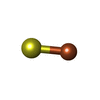
ChemComp-FES:
FE2/S2 (INORGANIC) CLUSTER / Iron–sulfur cluster

+
Macromolecule #8: IRON/SULFUR CLUSTER

MacromoleculeName: IRON/SULFUR CLUSTER / type: ligand / ID: 8 / Number of copies: 3 / Formula: SF4
Molecular weightTheoretical: 351.64 Da
Chemical component information

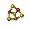
ChemComp-FS1:
IRON/SULFUR CLUSTER / Iron–sulfur cluster

+
Macromolecule #9: FE3-S4 CLUSTER

MacromoleculeName: FE3-S4 CLUSTER / type: ligand / ID: 9 / Number of copies: 3 / Formula: F3S
Molecular weightTheoretical: 295.795 Da
Chemical component information

ChemComp-F3S:
FE3-S4 CLUSTER / Iron–sulfur cluster

+
Macromolecule #10: 1,2-Distearoyl-sn-glycerophosphoethanolamine

MacromoleculeName: 1,2-Distearoyl-sn-glycerophosphoethanolamine / type: ligand / ID: 10 / Number of copies: 3 / Formula: 3PE
Molecular weightTheoretical: 748.065 Da
Chemical component information

ChemComp-3PE:
1,2-Distearoyl-sn-glycerophosphoethanolamine / phospholipid*YM / Phosphatidylethanolamine

+
Macromolecule #11: PROTOPORPHYRIN IX CONTAINING FE

MacromoleculeName: PROTOPORPHYRIN IX CONTAINING FE / type: ligand / ID: 11 / Number of copies: 3 / Formula: HEM
Molecular weightTheoretical: 616.487 Da
Chemical component information

ChemComp-HEM:
PROTOPORPHYRIN IX CONTAINING FE / Heme B

+
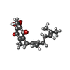
Macromolecule #12: UBIQUINONE-2

MacromoleculeName: UBIQUINONE-2 / type: ligand / ID: 12 / Number of copies: 3 / Formula: UQ2
Molecular weightTheoretical: 318.407 Da
Chemical component information

ChemComp-UQ2:
UBIQUINONE-2

-
Experimental details

-
Structure determination

Methodcryo EM
Processingsingle particle reconstruction
Aggregation stateparticle

-
Sample preparation

BufferpH: 7.5
VitrificationCryogen name: ETHANE

-
Electron microscopy

MicroscopeFEI TITAN KRIOS
Electron beamAcceleration voltage: 300 kV / Electron source: FIELD EMISSION GUN
Electron opticsIllumination mode: FLOOD BEAM / Imaging mode: BRIGHT FIELDBright-field microscopy
Image recordingFilm or detector model: GATAN K3 BIOQUANTUM (6k x 4k) / Average electron dose: 40.0 e/Å2
Experimental equipment
Model: Titan Krios / Image courtesy: FEI Company

-
Image processing

Startup modelType of model: NONE
Initial angle assignmentType: ANGULAR RECONSTITUTION
Final angle assignmentType: ANGULAR RECONSTITUTION
Final reconstructionResolution.type: BY AUTHOR / Resolution: 2.5 Å / Resolution method: FSC 0.143 CUT-OFF / Number images used: 38471

+
About Yorodumi

-
News

-
Feb 9, 2022. New format data for meta-information of EMDB entries

New format data for meta-information of EMDB entries

  • Version 3 of the EMDB header file is now the official format.
  • The previous official version 1.9 will be removed from the archive.

Related info.:EMDB header

External links:wwPDB to switch to version 3 of the EMDB data model

-
Aug 12, 2020. Covid-19 info

Covid-19 info

URL: https://pdbj.org/emnavi/covid19.php

New page: Covid-19 featured information page in EM Navigator.

Related info.:Covid-19 info / Mar 5, 2020. Novel coronavirus structure data

+
Mar 5, 2020. Novel coronavirus structure data

Novel coronavirus structure data

Related info.:Yorodumi Speices / Aug 12, 2020. Covid-19 info

External links:COVID-19 featured content - PDBj / Molecule of the Month (242):Coronavirus Proteases

+
Jan 31, 2019. EMDB accession codes are about to change! (news from PDBe EMDB page)

EMDB accession codes are about to change! (news from PDBe EMDB page)

  • The allocation of 4 digits for EMDB accession codes will soon come to an end. Whilst these codes will remain in use, new EMDB accession codes will include an additional digit and will expand incrementally as the available range of codes is exhausted. The current 4-digit format prefixed with “EMD-” (i.e. EMD-XXXX) will advance to a 5-digit format (i.e. EMD-XXXXX), and so on. It is currently estimated that the 4-digit codes will be depleted around Spring 2019, at which point the 5-digit format will come into force.
  • The EM Navigator/Yorodumi systems omit the EMD- prefix.

Related info.:Q: What is EMD? / ID/Accession-code notation in Yorodumi/EM Navigator

External links:EMDB Accession Codes are Changing Soon! / Contact to PDBj

+
Jul 12, 2017. Major update of PDB

Major update of PDB

  • wwPDB released updated PDB data conforming to the new PDBx/mmCIF dictionary.
  • This is a major update changing the version number from 4 to 5, and with Remediation, in which all the entries are updated.
  • In this update, many items about electron microscopy experimental information are reorganized (e.g. em_software).
  • Now, EM Navigator and Yorodumi are based on the updated data.

External links:wwPDB Remediation / Enriched Model Files Conforming to OneDep Data Standards Now Available in the PDB FTP Archive

-
Yorodumi

Thousand views of thousand structures

  • Yorodumi is a browser for structure data from EMDB, PDB, SASBDB, etc.
  • This page is also the successor to EM Navigator detail page, and also detail information page/front-end page for Omokage search.
  • The word "yorodu" (or yorozu) is an old Japanese word meaning "ten thousand". "mi" (miru) is to see.

Related info.:EMDB / PDB / SASBDB / Comparison of 3 databanks / Yorodumi Search / Aug 31, 2016. New EM Navigator & Yorodumi / Yorodumi Papers / Jmol/JSmol / Function and homology information / Changes in new EM Navigator and Yorodumi

Read more