 Movie
Movie Controller


+ Open data

- Basic information
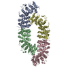
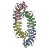
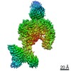
| Entry | Database: PDB / ID: 7fh1 | ||||||
|---|---|---|---|---|---|---|---|
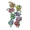
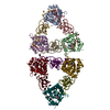
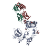
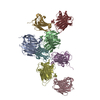
| Title | Structure of the human Meckelin | ||||||
 Components Components | Meckelin | ||||||
 Keywords Keywords | MEMBRANE PROTEIN / Cryo-EM | ||||||
| Function / homology |  Function and homology information Function and homology informationMKS complex / negative regulation of centrosome duplication / filamin binding / ciliary transition zone / non-canonical Wnt signaling pathway / ciliary membrane / cilium assembly / ERAD pathway / Anchoring of the basal body to the plasma membrane / cytoplasmic vesicle membrane ...MKS complex / negative regulation of centrosome duplication / filamin binding / ciliary transition zone / non-canonical Wnt signaling pathway / ciliary membrane / cilium assembly / ERAD pathway / Anchoring of the basal body to the plasma membrane / cytoplasmic vesicle membrane / unfolded protein binding / centrosome / endoplasmic reticulum membrane Similarity search - Function | ||||||
| Biological species |  Homo sapiens (human) Homo sapiens (human) | ||||||
| Method | ELECTRON MICROSCOPY / single particle reconstruction / cryo EM / Resolution: 3.34 Å | ||||||
 Authors Authors | Gong, D.S. | ||||||
| Funding support | 1items
| ||||||
 Citation Citation | Journal: Sci Adv / Year: 2021 Title: Structure of the human Meckel-Gruber protein Meckelin. Authors: Dongliang Liu / Dandan Qian / Huaizong Shen / Deshun Gong /  Abstract: Mutations in the gene account for most cases of the Meckel-Gruber syndrome, the most severe ciliopathy with a 100% mortality rate. Here, we report a 3.3-Å cryo–electron microscopy structure of ...Mutations in the gene account for most cases of the Meckel-Gruber syndrome, the most severe ciliopathy with a 100% mortality rate. Here, we report a 3.3-Å cryo–electron microscopy structure of human Meckelin (also known as TMEM67 and MKS3). The structure reveals a unique protein fold consisting of an unusual cysteine-rich domain that folds as an arch bridge stabilized by 11 pairs of disulfide bonds, a previously uncharacterized domain named β sheet–rich domain, a previously unidentified seven-transmembrane fold wherein TM4 to TM6 are broken near the cytoplasmic surface of the membrane, and a coiled-coil domain placed below the transmembrane domain. Meckelin forms a stable homodimer with an extensive dimer interface. Our structure establishes a framework for dissecting the function and disease mechanisms of Meckelin. | ||||||
| History |
|
- Structure visualization
| Movie |
Movie viewer |
|---|---|
| Structure viewer | Molecule: MolmilJmol/JSmol |
- Downloads & links
-Download
| PDBx/mmCIF format | 7fh1.cif.gz | 316.3 KB | Display | PDBx/mmCIF format |
|---|---|---|---|---|
| PDB format | pdb7fh1.ent.gz | 252.3 KB | Display | PDB format |
| PDBx/mmJSON format | 7fh1.json.gz | Tree view | PDBx/mmJSON format | |
| Others |  Other downloads Other downloads |
-Validation report
| Arichive directory | https://data.pdbj.org/pub/pdb/validation_reports/fh/7fh1ftp://data.pdbj.org/pub/pdb/validation_reports/fh/7fh1 | HTTPS FTP |
|---|
-Related structure data
| Related structure data | 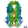 31584MC M: map data used to model this data C: citing same article ( |
|---|---|
| Similar structure data |

-Links
 PDBj
PDBj


- Assembly
| Deposited unit | 
|
|---|---|
| 1 |
|
-Components
| #1: Protein | Mass: 114980.438 Da / Num. of mol.: 2 Source method: isolated from a genetically manipulated source Source: (gene. exp.) Homo sapiens (human) / Gene: TMEM67, MKS3 / Production host: Homo sapiens (human) / References: UniProt: Q5HYA8#2: Polysaccharide | 2-acetamido-2-deoxy-beta-D-glucopyranose-(1-4)-2-acetamido-2-deoxy-beta-D-glucopyranose Source method: isolated from a genetically manipulated source #3: Polysaccharide | Source method: isolated from a genetically manipulated source Has ligand of interest | N | Has protein modification | Y | |
|---|
-Experimental details
-Experiment
| Experiment | Method: ELECTRON MICROSCOPY |
|---|---|
| EM experiment | Aggregation state: PARTICLE / 3D reconstruction method: single particle reconstruction |
- Sample preparation
| Component | Name: homodimer of Meckelin / Type: COMPLEX / Entity ID: #1 / Source: RECOMBINANT |
|---|---|
| Source (natural) | Organism: Homo sapiens (human) |
| Source (recombinant) | Organism: Homo sapiens (human) |
| Buffer solution | pH: 8 |
| Specimen | Embedding applied: NO / Shadowing applied: NO / Staining applied: NO / Vitrification applied: YES |
| Vitrification | Cryogen name: ETHANE |
- Electron microscopy imaging
| Experimental equipment |  Model: Titan Krios / Image courtesy: FEI Company |
|---|---|
| Microscopy | Model: FEI TITAN KRIOS |
| Electron gun | Electron source:  FIELD EMISSION GUN / Accelerating voltage: 300 kV / Illumination mode: FLOOD BEAM FIELD EMISSION GUN / Accelerating voltage: 300 kV / Illumination mode: FLOOD BEAM |
| Electron lens | Mode: BRIGHT FIELD |
| Image recording | Electron dose: 50 e/Å2 / Film or detector model: GATAN K3 (6k x 4k) |
- Processing
| CTF correction | Type: PHASE FLIPPING AND AMPLITUDE CORRECTION |
|---|---|
| 3D reconstruction | Resolution: 3.34 Å / Resolution method: FSC 0.143 CUT-OFF / Num. of particles: 219967 / Symmetry type: POINT |
