 Movie
Movie Controller


+ Open data

- Basic information
| Entry | Database: EMDB / ID: EMD-31584 | |||||||||
|---|---|---|---|---|---|---|---|---|---|---|
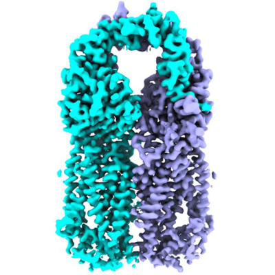
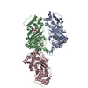
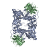
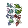
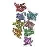
| Title | Structure of the human Meckelin | |||||||||
 Map data Map data | ||||||||||
 Sample Sample |
| |||||||||
 Keywords Keywords | Cryo-EM / MEMBRANE PROTEIN | |||||||||
| Function / homology |  Function and homology information Function and homology informationMKS complex / negative regulation of centrosome duplication / filamin binding / ciliary transition zone / non-canonical Wnt signaling pathway / ciliary membrane / cilium assembly / ERAD pathway / Anchoring of the basal body to the plasma membrane / cytoplasmic vesicle membrane ...MKS complex / negative regulation of centrosome duplication / filamin binding / ciliary transition zone / non-canonical Wnt signaling pathway / ciliary membrane / cilium assembly / ERAD pathway / Anchoring of the basal body to the plasma membrane / cytoplasmic vesicle membrane / unfolded protein binding / centrosome / endoplasmic reticulum membrane Similarity search - Function | |||||||||
| Biological species |  Homo sapiens (human) Homo sapiens (human) | |||||||||
| Method | single particle reconstruction / cryo EM / Resolution: 3.34 Å | |||||||||
 Authors Authors | Gong DS | |||||||||
| Funding support | 1 items
| |||||||||
 Citation Citation | Journal: Sci Adv / Year: 2021 Title: Structure of the human Meckel-Gruber protein Meckelin. Authors: Dongliang Liu / Dandan Qian / Huaizong Shen / Deshun Gong /  Abstract: Mutations in the gene account for most cases of the Meckel-Gruber syndrome, the most severe ciliopathy with a 100% mortality rate. Here, we report a 3.3-Å cryo–electron microscopy structure of ...Mutations in the gene account for most cases of the Meckel-Gruber syndrome, the most severe ciliopathy with a 100% mortality rate. Here, we report a 3.3-Å cryo–electron microscopy structure of human Meckelin (also known as TMEM67 and MKS3). The structure reveals a unique protein fold consisting of an unusual cysteine-rich domain that folds as an arch bridge stabilized by 11 pairs of disulfide bonds, a previously uncharacterized domain named β sheet–rich domain, a previously unidentified seven-transmembrane fold wherein TM4 to TM6 are broken near the cytoplasmic surface of the membrane, and a coiled-coil domain placed below the transmembrane domain. Meckelin forms a stable homodimer with an extensive dimer interface. Our structure establishes a framework for dissecting the function and disease mechanisms of Meckelin. | |||||||||
| History |
|
- Structure visualization
| Movie |
Movie viewer |
|---|---|
| Structure viewer | EM map: SurfViewMolmilJmol/JSmol |
| Supplemental images |
- Downloads & links
-EMDB archive
| Map data | emd_31584.map.gz | 49.1 MB | EMDB map data format | |
|---|---|---|---|---|
| Header (meta data) | emd-31584-v30.xmlemd-31584.xml | 9.2 KB 9.2 KB | Display Display | EMDB header |
| FSC (resolution estimation) | emd_31584_fsc.xml | 8.6 KB | Display | FSC data file |
| Images |  emd_31584.png emd_31584.png | 183.4 KB | ||
| Filedesc metadata | emd-31584.cif.gz | 5.5 KB | ||
| Archive directory |  http://ftp.pdbj.org/pub/emdb/structures/EMD-31584ftp://ftp.pdbj.org/pub/emdb/structures/EMD-31584 http://ftp.pdbj.org/pub/emdb/structures/EMD-31584ftp://ftp.pdbj.org/pub/emdb/structures/EMD-31584 | HTTPS FTP |
-Validation report
| Summary document | emd_31584_validation.pdf.gz | 520.4 KB | Display | EMDB validaton report |
|---|---|---|---|---|
| Full document | emd_31584_full_validation.pdf.gz | 520 KB | Display | |
| Data in XML | emd_31584_validation.xml.gz | 10.5 KB | Display | |
| Data in CIF | emd_31584_validation.cif.gz | 13.8 KB | Display | |
| Arichive directory | https://ftp.pdbj.org/pub/emdb/validation_reports/EMD-31584ftp://ftp.pdbj.org/pub/emdb/validation_reports/EMD-31584 | HTTPS FTP |
-Related structure data
| Related structure data |  7fh1MC M: atomic model generated by this map C: citing same article ( |
|---|---|
| Similar structure data |


-Links
| EMDB pages | EMDB (EBI/PDBe) / EMDataResource |
|---|---|
| Related items in Molecule of the Month |


-Map
| File | Download / File: emd_31584.map.gz / Format: CCP4 / Size: 52.7 MB / Type: IMAGE STORED AS FLOATING POINT NUMBER (4 BYTES) | ||||||||||||||||||||||||||||||||||||||||||||||||||||||||||||||||||||
|---|---|---|---|---|---|---|---|---|---|---|---|---|---|---|---|---|---|---|---|---|---|---|---|---|---|---|---|---|---|---|---|---|---|---|---|---|---|---|---|---|---|---|---|---|---|---|---|---|---|---|---|---|---|---|---|---|---|---|---|---|---|---|---|---|---|---|---|---|---|
| Projections & slices | Image control
Images are generated by Spider. | ||||||||||||||||||||||||||||||||||||||||||||||||||||||||||||||||||||
| Voxel size | X=Y=Z: 1.087 Å | ||||||||||||||||||||||||||||||||||||||||||||||||||||||||||||||||||||
| Density |
| ||||||||||||||||||||||||||||||||||||||||||||||||||||||||||||||||||||
| Symmetry | Space group: 1 | ||||||||||||||||||||||||||||||||||||||||||||||||||||||||||||||||||||
| Details | EMDB XML:
CCP4 map header:
| ||||||||||||||||||||||||||||||||||||||||||||||||||||||||||||||||||||
 Z (Sec.)
 Y (Row.)
 X (Col.)


-Supplemental data
- Sample components
-Entire : homodimer of Meckelin
| Entire | Name: homodimer of Meckelin |
|---|---|
| Components |
|
-Supramolecule #1: homodimer of Meckelin
| Supramolecule | Name: homodimer of Meckelin / type: complex / ID: 1 / Parent: 0 / Macromolecule list: all |
|---|---|
| Source (natural) | Organism: Homo sapiens (human) |
-Macromolecule #1: Meckelin
| Macromolecule | Name: Meckelin / type: protein_or_peptide / ID: 1 / Number of copies: 2 / Enantiomer: LEVO |
|---|---|
| Source (natural) | Organism: Homo sapiens (human) |
| Molecular weight | Theoretical: 114.980438 KDa |
| Recombinant expression | Organism: Homo sapiens (human) |
| Sequence | String: MATRGGAGVA MAVWSLLSAR AVTAFLLLFL PRFLQAQTFS FPFQQPEKCD NNQYFDISAL SCVPCGANQR QDARGTSCVC LPGFQMISN NGGPAIICKK CPENMKGVTE DGWNCISCPS DLTAEGKCHC PIGHILVERD INGTLLSQAT CELCDGNENS F MVVNALGD ...String: MATRGGAGVA MAVWSLLSAR AVTAFLLLFL PRFLQAQTFS FPFQQPEKCD NNQYFDISAL SCVPCGANQR QDARGTSCVC LPGFQMISN NGGPAIICKK CPENMKGVTE DGWNCISCPS DLTAEGKCHC PIGHILVERD INGTLLSQAT CELCDGNENS F MVVNALGD RCVRCEPTFV NTSRSCACSE PNILTGGLCF SSTGNFPLRR ISAARYGEVG MSLTSEWFAK YLQSSAAACW VY ANLTSCQ ALGNMCVMNM NSYDFATFDA CGLFQFIFEN TAGLSTVHSI SFWRQNLPWL FYGDQLGLAP QVLSSTSLPT NFS FKGENQ NTKLKFVAAS YDIRGNFLKW QTLEGGVLQL CPDTETRLNA AYSFGTTYQQ NCEIPISKIL IDFPTPIFYD VYLE YTDEN QHQYILAVPV LNLNLQHNKI FVNQDSNSGK WLLTRRIFLV DAVSGRENDL GTQPRVIRVA TQISLSVHLV PNTIN GNIY PPLITIAYSD IDIKDANSQS VKVSFSVTYE MDHGEAHVQT DIALGVLGGL AVLASLLKTA GWKRRIGSPM IDLQTV VKF LVYYAGDLAN VFFIITVGTG LYWLIFFKAQ KSVSVLLPMP IQEERFVTYV GCAFALKALQ FLHKLISQIT IDVFFID WE RPKGKVLKAV EGEGGVRSAT VPVSIWRTYF VANEWNEIQT VRKINSLFQV LTVLFFLEVV GFKNLALMDS SSSLSRNP P SYIAPYSCIL RYAVSAALWL AIGIIQVVFF AVFYERFIED KIRQFVDLCS MSNISVFLLS HKCFGYYIHG RSVHGHADT NMEEMNMNLK REAENLCSQR GLVPNTDGQT FEIAISNQMR QHYDRIHETL IRKNGPARLL SSSASTFEQS IKAYHMMNKF LGSFIDHVH KEMDYFIKDK LLLERILGME FMEPMEKSIF YNDEGYSFSS VLYYGNEATL LIFDLLFFCV VDLACQNFIL A SFLTYLQQ EIFRYIRNTV GQKNLASKTL VDQRFLILEG SHHHHHHHHH HGSVEDYKDD DDK UniProtKB: Meckelin |
-Experimental details
-Structure determination
| Method | cryo EM |
|---|---|
 Processing Processing | single particle reconstruction |
| Aggregation state | particle |
-Sample preparation
| Buffer | pH: 8 |
|---|---|
| Vitrification | Cryogen name: ETHANE |
- Electron microscopy
| Microscope | FEI TITAN KRIOS |
|---|---|
| Image recording | Film or detector model: GATAN K3 (6k x 4k) / Average electron dose: 50.0 e/Å2 |
| Electron beam | Acceleration voltage: 300 kV / Electron source:  FIELD EMISSION GUN FIELD EMISSION GUN |
| Electron optics | Illumination mode: FLOOD BEAM / Imaging mode: BRIGHT FIELD |
| Experimental equipment |  Model: Titan Krios / Image courtesy: FEI Company |

