 Movie
Movie Controller

[English] 日本語
 Yorodumi
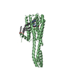
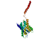
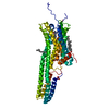
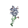
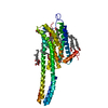
- PDB-7f2f: The complex of DNA with the C-terminal domain of TYE7 from Saccha... -
+ Open data

- Basic information
| Entry | Database: PDB / ID: 7f2f | ||||||
|---|---|---|---|---|---|---|---|
| Title | The complex of DNA with the C-terminal domain of TYE7 from Saccharomyces cerevisiae. | ||||||
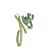
 Components Components |
| ||||||
 Keywords Keywords | TRANSCRIPTION / Glycolysis / bHLH transcription factor | ||||||
| Function / homology |  Function and homology information Function and homology informationDNA-binding transcription activator activity / positive regulation of glycolytic process / transcription elongation by RNA polymerase II / sequence-specific DNA binding / protein dimerization activity / RNA polymerase II cis-regulatory region sequence-specific DNA binding / DNA-binding transcription factor activity / chromatin / positive regulation of transcription by RNA polymerase II / nucleus Similarity search - Function | ||||||
| Biological species |  synthetic construct (others) | ||||||
| Method |  X-RAY DIFFRACTION / SYNCHROTRON / MOLECULAR REPLACEMENT / molecular replacement / Resolution: 2.55 Å X-RAY DIFFRACTION / SYNCHROTRON / MOLECULAR REPLACEMENT / molecular replacement / Resolution: 2.55 Å | ||||||
 Authors Authors | Gui, W. | ||||||
| Funding support | 1items
| ||||||
 Citation Citation | Journal: Acta Crystallogr.,Sect.F / Year: 2021 Title: Crystal structure of the complex of DNA with the C-terminal domain of TYE7 from Saccharomyces cerevisiae. Authors: Gui, W. / Xue, L. / Yue, J. / Kuang, Z. / Jin, Y. / Niu, L. | ||||||
| History |
|
- Structure visualization
| Structure viewer | Molecule: MolmilJmol/JSmol |
|---|
- Downloads & links
-Download
| PDBx/mmCIF format | 7f2f.cif.gz | 73 KB | Display | PDBx/mmCIF format |
|---|---|---|---|---|
| PDB format | pdb7f2f.ent.gz | 49.4 KB | Display | PDB format |
| PDBx/mmJSON format | 7f2f.json.gz | Tree view | PDBx/mmJSON format | |
| Others |  Other downloads Other downloads |
-Validation report
| Summary document | 7f2f_validation.pdf.gz | 459.1 KB | Display | wwPDB validaton report |
|---|---|---|---|---|
| Full document | 7f2f_full_validation.pdf.gz | 461.5 KB | Display | |
| Data in XML | 7f2f_validation.xml.gz | 9.5 KB | Display | |
| Data in CIF | 7f2f_validation.cif.gz | 12.1 KB | Display | |
| Arichive directory | https://data.pdbj.org/pub/pdb/validation_reports/f2/7f2fftp://data.pdbj.org/pub/pdb/validation_reports/f2/7f2f | HTTPS FTP |
-Related structure data
| Related structure data |  1am9S S: Starting model for refinement |
|---|---|
| Similar structure data |


-Links
 PDBj
PDBj


- Assembly
| Deposited unit | 
| ||||||||
|---|---|---|---|---|---|---|---|---|---|
| 1 |
| ||||||||
| Unit cell |
| ||||||||
| Components on special symmetry positions |
|
-Components
| #1: Protein | Mass: 15858.110 Da / Num. of mol.: 2 Source method: isolated from a genetically manipulated source Source: (gene. exp.) Gene: TYE7, SGC1, YOR344C, O6233 / Production host:  #2: DNA chain | | Mass: 4584.984 Da / Num. of mol.: 1 / Source method: obtained synthetically / Source: (synth.) synthetic construct (others) #3: DNA chain | | Mass: 4593.998 Da / Num. of mol.: 1 / Source method: obtained synthetically / Source: (synth.) synthetic construct (others) |
|---|
-Experimental details
-Experiment
| Experiment | Method: X-RAY DIFFRACTION / Number of used crystals: 1 |
|---|
- Sample preparation
| Crystal | Density Matthews: 2.96 Å3/Da / Density % sol: 58.49 % |
|---|---|
| Crystal grow | Temperature: 283 K / Method: vapor diffusion, sitting drop Details: 0.1M HEPES pH7.5, 10% w/v PEG 8000, 11% v/v ethylene glycol |
-Data collection
| Diffraction | Mean temperature: 100 K / Serial crystal experiment: N |
|---|---|
| Diffraction source | Source: SYNCHROTRON / Site: SSRF  / Beamline: BL18U1 / Wavelength: 0.97853 Å / Beamline: BL18U1 / Wavelength: 0.97853 Å |
| Detector | Type: DECTRIS PILATUS 6M / Detector: PIXEL / Date: May 4, 2019 |
| Radiation | Protocol: SINGLE WAVELENGTH / Monochromatic (M) / Laue (L): M / Scattering type: x-ray |
| Radiation wavelength | Wavelength: 0.97853 Å / Relative weight: 1 |
| Reflection | Resolution: 2.55→67.76 Å / Num. obs: 16802 / % possible obs: 99.9 % / Redundancy: 17.9 % / CC1/2: 0.995 / CC star: 0.999 / Net I/σ(I): 33.15 |
| Reflection shell | Resolution: 2.55→2.59 Å / Mean I/σ(I) obs: 2.05 / Num. unique obs: 818 / CC1/2: 0.876 / CC star: 0.967 |
-Phasing
| Phasing | Method: molecular replacement |
|---|
- Processing
| Software |
| ||||||||||||||||||||||||||||||||||||||||||||||||||||||||||||
|---|---|---|---|---|---|---|---|---|---|---|---|---|---|---|---|---|---|---|---|---|---|---|---|---|---|---|---|---|---|---|---|---|---|---|---|---|---|---|---|---|---|---|---|---|---|---|---|---|---|---|---|---|---|---|---|---|---|---|---|---|---|
| Refinement | Method to determine structure: MOLECULAR REPLACEMENT Starting model: 1AM9 Resolution: 2.55→67.76 Å / Cor.coef. Fo:Fc: 0.924 / Cor.coef. Fo:Fc free: 0.897 / SU B: 10.87 / SU ML: 0.229 / Cross valid method: THROUGHOUT / σ(F): 0 / ESU R: 0.352 / ESU R Free: 0.273 / Stereochemistry target values: MAXIMUM LIKELIHOOD Details: HYDROGENS HAVE BEEN ADDED IN THE RIDING POSITIONS U VALUES : REFINED INDIVIDUALLY
| ||||||||||||||||||||||||||||||||||||||||||||||||||||||||||||
| Solvent computation | Ion probe radii: 0.8 Å / Shrinkage radii: 0.8 Å / VDW probe radii: 1.2 Å / Solvent model: MASK | ||||||||||||||||||||||||||||||||||||||||||||||||||||||||||||
| Displacement parameters | Biso max: 190.25 Å2 / Biso mean: 70.705 Å2 / Biso min: 28.75 Å2
| ||||||||||||||||||||||||||||||||||||||||||||||||||||||||||||
| Refinement step | Cycle: final / Resolution: 2.55→67.76 Å
| ||||||||||||||||||||||||||||||||||||||||||||||||||||||||||||
| Refine LS restraints |
| ||||||||||||||||||||||||||||||||||||||||||||||||||||||||||||
| LS refinement shell | Resolution: 2.551→2.617 Å / Rfactor Rfree error: 0 / Total num. of bins used: 20
|
