[English] 日本語
Yorodumi
- PDB-4oj2: The structure of aquaporin -

+
Open data


ID or keywords:

Loading...

-
Basic information

Entry
Database: PDB / ID: 4oj2
TitleThe structure of aquaporin
ComponentsAquaporin-2
KeywordsTRANSPORT PROTEIN / aquaporin-2 / water transport simulations / c-terminal alpha helix / Structural Genomics / Protein Structure Initiative / Transcontinental EM Initiative for Membrane Protein Structure / TEMIMPS / NPA motif / Water Channel / Membrane / PSI-Biology
Function / homology
Function and homology information


cellular response to water deprivation / renal water transport / glycerol transmembrane transporter activity / Passive transport by Aquaporins / glycerol transmembrane transport / lumenal side of membrane / water transmembrane transporter activity / cellular response to mercury ion / water transport / water channel activity ...cellular response to water deprivation / renal water transport / glycerol transmembrane transporter activity / Passive transport by Aquaporins / glycerol transmembrane transport / lumenal side of membrane / water transmembrane transporter activity / cellular response to mercury ion / water transport / water channel activity / metanephric collecting duct development / renal water homeostasis / transport vesicle membrane / cellular response to copper ion / actin filament organization / recycling endosome / Vasopressin regulates renal water homeostasis via Aquaporins / basolateral plasma membrane / protein homotetramerization / apical plasma membrane / perinuclear region of cytoplasm / Golgi apparatus / extracellular exosome / membrane / plasma membrane
Similarity search - Function
Glycerol uptake facilitator protein / Glycerol uptake facilitator protein. / Aquaporin transporter / Major intrinsic protein, conserved site / MIP family signature. / Major intrinsic protein / Major intrinsic protein / Aquaporin-like / Up-down Bundle / Mainly Alpha
Similarity search - Domain/homology
Biological speciesHomo sapiens (human)
MethodX-RAY DIFFRACTION / SYNCHROTRON / MOLECULAR REPLACEMENT / Resolution: 3.05 Å
AuthorsVahedi-Faridi, A. / Lodowski, D. / Engel, A. / Transcontinental EM Initiative for Membrane Protein Structure (TEMIMPS)
CitationJournal: To be Published
Title: The structure of aquaporin
Authors: Vahedi-Faridi, A. / Lodowski, D. / Schenk, A. / Kaptan, S. / De groot, B. / Walz, T. / Engel, A.
History
DepositionJan 20, 2014Deposition site: RCSB / Processing site: RCSB
Revision 1.0May 7, 2014Provider: repository / Type: Initial release
Revision 1.1Feb 28, 2024Group: Data collection / Database references
Category: chem_comp_atom / chem_comp_bond ...chem_comp_atom / chem_comp_bond / database_2 / struct_ref_seq_dif
Item: _database_2.pdbx_DOI / _database_2.pdbx_database_accession / _struct_ref_seq_dif.details

-
Structure visualization

Structure viewerMolecule:
MolmilJmol/JSmol

Downloads & links

-
Assembly

Deposited unit
X: Aquaporin-2


Theoretical massNumber of molelcules
Total (without water)29,1711
Polymers29,1711
Non-polymers00
Water45025
1
X: Aquaporin-2

X: Aquaporin-2

X: Aquaporin-2

X: Aquaporin-2


Theoretical massNumber of molelcules
Total (without water)116,6834
Polymers116,6834
Non-polymers00
Water724
TypeNameSymmetry operationNumber
identity operation1_555x,y,z1
crystal symmetry operation2_565-x,-y+1,z1
crystal symmetry operation3_555-y+1/2,x+1/2,z1
crystal symmetry operation4_455y-1/2,-x+1/2,z1
Buried area13540 Å2
ΔGint-141 kcal/mol
Surface area40470 Å2
MethodPISA
Unit cell
Length a, b, c (Å)95.717, 95.717, 79.058
Angle α, β, γ (deg.)90.00, 90.00, 90.00
Int Tables number90
Space group name H-MP4212

-
Components

#1: Protein 
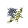
Aquaporin-2 / AQP-2 / ADH water channel / Aquaporin-CD / AQP-CD / Collecting duct water channel protein / WCH-CD ...AQP-2 / ADH water channel / Aquaporin-CD / AQP-CD / Collecting duct water channel protein / WCH-CD / Water channel protein for renal collecting duct


Mass: 29170.740 Da / Num. of mol.: 1 / Mutation: S256A
Source method: isolated from a genetically manipulated source
Source: (gene. exp.) Homo sapiens (human) / Gene: AQP2 / Production host: 
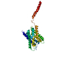
Spodoptera frugiperda (fall armyworm) / Strain (production host): Sf9 / References: UniProt: P41181
#2: Water ChemComp-HOH / water


Mass: 18.015 Da / Num. of mol.: 25 / Source method: isolated from a natural source / Formula: H2O

-
Experimental details

-
Experiment

ExperimentMethod: X-RAY DIFFRACTION / Number of used crystals: 1

-
Sample preparation

CrystalDensity Matthews: 3.1 Å3/Da / Density % sol: 60.37 %
Crystal growTemperature: 298 K / Method: vapor diffusion, sitting drop / pH: 7
Details: 30% PEG 400, 0.1M sodium chloride, 0.1M MOPS, pH 7.0, VAPOR DIFFUSION, SITTING DROP, temperature 298.0K

-
Data collection

DiffractionMean temperature: 100 K
Diffraction sourceSource: SYNCHROTRON / Site: APS / Beamline: 24-ID-C / Wavelength: 0.9792 Å
DetectorType: ADSC QUANTUM 315r / Detector: CCD / Date: Aug 8, 2013
RadiationMonochromator: double crystal / Protocol: SINGLE WAVELENGTH / Monochromatic (M) / Laue (L): M / Scattering type: x-ray
Radiation wavelengthWavelength: 0.9792 Å / Relative weight: 1
ReflectionResolution: 3.05→79.07 Å / Num. all: 7130 / Num. obs: 7153 / % possible obs: 99.68 % / Observed criterion σ(F): 1 / Observed criterion σ(I): 1 / Redundancy: 13.8 % / Net I/σ(I): 38.8
Reflection shellResolution: 3.05→3.29 Å / % possible all: 100

-
Processing

Software
NameVersionClassification
ADSCQuantumdata collection
SOLVEphasing
REFMAC5.8.0049refinement
HKL-2000data reduction
HKL-2000data scaling
RefinementMethod to determine structure: MOLECULAR REPLACEMENT / Resolution: 3.05→79.07 Å / Cor.coef. Fo:Fc: 0.924 / Cor.coef. Fo:Fc free: 0.895 / SU B: 56.229 / SU ML: 0.465 / Cross valid method: THROUGHOUT / ESU R Free: 0.479 / Stereochemistry target values: MAXIMUM LIKELIHOOD / Details: HYDROGENS HAVE BEEN ADDED IN THE RIDING POSITIONS
RfactorNum. reflection% reflectionSelection details
Rfree0.29897 300 4 %RANDOM
Rwork0.25707 ---
obs0.25893 7130 99.68 %-
all-7153 --
Solvent computationIon probe radii: 0.8 Å / Shrinkage radii: 0.8 Å / VDW probe radii: 1.2 Å / Solvent model: MASK
Displacement parametersBiso mean: 150.323 Å2
Baniso -1Baniso -2Baniso -3
1--2.58 Å20 Å20 Å2
2---2.58 Å20 Å2
3---5.15 Å2
Refinement stepCycle: LAST / Resolution: 3.05→79.07 Å
ProteinNucleic acidLigandSolventTotal
Num. atoms1890 0 0 25 1915
Refine LS restraints
Refine-IDTypeDev idealDev ideal targetNumber
X-RAY DIFFRACTIONr_bond_refined_d0.0030.0191935
X-RAY DIFFRACTIONr_bond_other_d00.021898
X-RAY DIFFRACTIONr_angle_refined_deg0.6931.9612643
X-RAY DIFFRACTIONr_angle_other_deg0.49834332
X-RAY DIFFRACTIONr_dihedral_angle_1_deg4.4855252
X-RAY DIFFRACTIONr_dihedral_angle_2_deg31.36722.63972
X-RAY DIFFRACTIONr_dihedral_angle_3_deg16.98115287
X-RAY DIFFRACTIONr_dihedral_angle_4_deg14.0571512
X-RAY DIFFRACTIONr_chiral_restr0.0380.2316
X-RAY DIFFRACTIONr_gen_planes_refined0.0040.0212200
X-RAY DIFFRACTIONr_gen_planes_other00.02454
X-RAY DIFFRACTIONr_nbd_refined
X-RAY DIFFRACTIONr_nbd_other
X-RAY DIFFRACTIONr_nbtor_refined
X-RAY DIFFRACTIONr_nbtor_other
X-RAY DIFFRACTIONr_xyhbond_nbd_refined
X-RAY DIFFRACTIONr_xyhbond_nbd_other
X-RAY DIFFRACTIONr_metal_ion_refined
X-RAY DIFFRACTIONr_metal_ion_other
X-RAY DIFFRACTIONr_symmetry_vdw_refined
X-RAY DIFFRACTIONr_symmetry_vdw_other
X-RAY DIFFRACTIONr_symmetry_hbond_refined
X-RAY DIFFRACTIONr_symmetry_hbond_other
X-RAY DIFFRACTIONr_symmetry_metal_ion_refined
X-RAY DIFFRACTIONr_symmetry_metal_ion_other
X-RAY DIFFRACTIONr_mcbond_it2.7449.6621011
X-RAY DIFFRACTIONr_mcbond_other2.7459.6561010
X-RAY DIFFRACTIONr_mcangle_it4.40614.4751262
X-RAY DIFFRACTIONr_mcangle_other4.40414.4821263
X-RAY DIFFRACTIONr_scbond_it2.2819.934924
X-RAY DIFFRACTIONr_scbond_other2.289.939925
X-RAY DIFFRACTIONr_scangle_it
X-RAY DIFFRACTIONr_scangle_other3.79214.811382
X-RAY DIFFRACTIONr_long_range_B_refined7.08181.0242558
X-RAY DIFFRACTIONr_long_range_B_other7.0881.0512559
X-RAY DIFFRACTIONr_rigid_bond_restr
X-RAY DIFFRACTIONr_sphericity_free
X-RAY DIFFRACTIONr_sphericity_bonded
LS refinement shellResolution: 3.048→3.127 Å / Total num. of bins used: 20
RfactorNum. reflection% reflection
Rfree0.398 32 -
Rwork0.34 499 -
obs--99.81 %
Refinement TLS params.

Method: refined / Refine-ID: X-RAY DIFFRACTION

IDL112)L122)L132)L222)L232)L332)S11 (Å °)S12 (Å °)S13 (Å °)S21 (Å °)S22 (Å °)S23 (Å °)S31 (Å °)S32 (Å °)S33 (Å °)T112)T122)T132)T222)T232)T332)Origin x (Å)Origin y (Å)Origin z (Å)
14.34030.1731-0.58734.17691.25311.46240.0243-0.0346-0.66560.13820.02190.39660.4562-0.2682-0.04610.3771-0.0494-0.06920.32020.12060.2244-9.249530.58424.024
29.5121-3.20167.80891.1322-2.56866.9311-0.15670.3668-0.74630.04480.06080.451-0.5538-0.00910.09591.4962-0.0388-0.15481.4089-0.03741.4025-38.877338.9374-11.697
33.2747-1.8382-0.64131.23070.92481.8211-0.09580.0526-0.63870.1149-0.18020.40940.2986-0.2680.2760.5058-0.12460.06740.4028-0.06760.2464-8.990329.962223.9785
Refinement TLS group
IDRefine-IDRefine TLS-IDAuth asym-IDAuth seq-ID
1X-RAY DIFFRACTION1X5 - 31
2X-RAY DIFFRACTION1X32 - 41
3X-RAY DIFFRACTION1X42 - 108
4X-RAY DIFFRACTION1X109 - 126
5X-RAY DIFFRACTION1X127 - 149
6X-RAY DIFFRACTION1X150 - 160
7X-RAY DIFFRACTION1X161 - 221
8X-RAY DIFFRACTION2X222 - 233
9X-RAY DIFFRACTION2X234 - 257
10X-RAY DIFFRACTION3X301 - 325

+
About Yorodumi

-
News

-
Feb 9, 2022. New format data for meta-information of EMDB entries

New format data for meta-information of EMDB entries

  • Version 3 of the EMDB header file is now the official format.
  • The previous official version 1.9 will be removed from the archive.

Related info.:EMDB header

External links:wwPDB to switch to version 3 of the EMDB data model

-
Aug 12, 2020. Covid-19 info

Covid-19 info

URL: https://pdbjlvh1.pdbj.org/emnavi/covid19.php

New page: Covid-19 featured information page in EM Navigator.

Related info.:Covid-19 info / Mar 5, 2020. Novel coronavirus structure data

+
Mar 5, 2020. Novel coronavirus structure data

Novel coronavirus structure data

Related info.:Yorodumi Speices / Aug 12, 2020. Covid-19 info

External links:COVID-19 featured content - PDBj / Molecule of the Month (242):Coronavirus Proteases

+
Jan 31, 2019. EMDB accession codes are about to change! (news from PDBe EMDB page)

EMDB accession codes are about to change! (news from PDBe EMDB page)

  • The allocation of 4 digits for EMDB accession codes will soon come to an end. Whilst these codes will remain in use, new EMDB accession codes will include an additional digit and will expand incrementally as the available range of codes is exhausted. The current 4-digit format prefixed with “EMD-” (i.e. EMD-XXXX) will advance to a 5-digit format (i.e. EMD-XXXXX), and so on. It is currently estimated that the 4-digit codes will be depleted around Spring 2019, at which point the 5-digit format will come into force.
  • The EM Navigator/Yorodumi systems omit the EMD- prefix.

Related info.:Q: What is EMD? / ID/Accession-code notation in Yorodumi/EM Navigator

External links:EMDB Accession Codes are Changing Soon! / Contact to PDBj

+
Jul 12, 2017. Major update of PDB

Major update of PDB

  • wwPDB released updated PDB data conforming to the new PDBx/mmCIF dictionary.
  • This is a major update changing the version number from 4 to 5, and with Remediation, in which all the entries are updated.
  • In this update, many items about electron microscopy experimental information are reorganized (e.g. em_software).
  • Now, EM Navigator and Yorodumi are based on the updated data.

External links:wwPDB Remediation / Enriched Model Files Conforming to OneDep Data Standards Now Available in the PDB FTP Archive

-
Yorodumi

Thousand views of thousand structures

  • Yorodumi is a browser for structure data from EMDB, PDB, SASBDB, etc.
  • This page is also the successor to EM Navigator detail page, and also detail information page/front-end page for Omokage search.
  • The word "yorodu" (or yorozu) is an old Japanese word meaning "ten thousand". "mi" (miru) is to see.

Related info.:EMDB / PDB / SASBDB / Comparison of 3 databanks / Yorodumi Search / Aug 31, 2016. New EM Navigator & Yorodumi / Yorodumi Papers / Jmol/JSmol / Function and homology information / Changes in new EM Navigator and Yorodumi

Read more