 Movie
Movie Controller

[English] 日本語
 Yorodumi
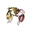
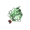
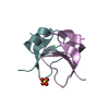
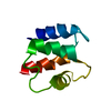
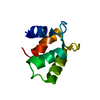
- PDB-7dvc: Crystal structure of the computationally designed reDPBB_sym1 protein -
+ Open data

- Basic information
| Entry | Database: PDB / ID: 7dvc | ||||||
|---|---|---|---|---|---|---|---|
| Title | Crystal structure of the computationally designed reDPBB_sym1 protein | ||||||
 Components Components | reDPBB_sym1 protein | ||||||
 Keywords Keywords | CHAPERONE / Double psi beta barrel | ||||||
| Function / homology | Barwin-like endoglucanases - #20 / Barwin-like endoglucanases / Beta Barrel / Mainly Beta / ACETATE ION Function and homology information Function and homology information | ||||||
| Biological species | synthetic construct (others) | ||||||
| Method |  X-RAY DIFFRACTION / SYNCHROTRON / MOLECULAR REPLACEMENT / Resolution: 1.705 Å X-RAY DIFFRACTION / SYNCHROTRON / MOLECULAR REPLACEMENT / Resolution: 1.705 Å | ||||||
 Authors Authors | Yagi, S. / Tagami, S. / Padhi, A.K. / Zhang, K.Y.J. | ||||||
| Funding support |  Japan, 1items Japan, 1items
| ||||||
 Citation Citation | Journal: J.Am.Chem.Soc. / Year: 2021 Title: Seven Amino Acid Types Suffice to Create the Core Fold of RNA Polymerase. Authors: Yagi, S. / Padhi, A.K. / Vucinic, J. / Barbe, S. / Schiex, T. / Nakagawa, R. / Simoncini, D. / Zhang, K.Y.J. / Tagami, S. | ||||||
| History |
|
- Structure visualization
| Structure viewer | Molecule: MolmilJmol/JSmol |
|---|
- Downloads & links
-Download
| PDBx/mmCIF format | 7dvc.cif.gz | 229.5 KB | Display | PDBx/mmCIF format |
|---|---|---|---|---|
| PDB format | pdb7dvc.ent.gz | 187.7 KB | Display | PDB format |
| PDBx/mmJSON format | 7dvc.json.gz | Tree view | PDBx/mmJSON format | |
| Others |  Other downloads Other downloads |
-Validation report
| Arichive directory | https://data.pdbj.org/pub/pdb/validation_reports/dv/7dvcftp://data.pdbj.org/pub/pdb/validation_reports/dv/7dvc | HTTPS FTP |
|---|
-Related structure data
| Related structure data |  7dboC  7dg7C 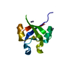 7dg9C 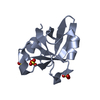 7di0C 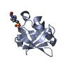 7di1C  7du6C 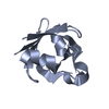 7du7C 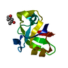 7dvfC 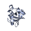 7dvhC  7dwwC 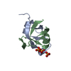 7dxrC 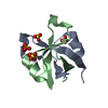 7dxsC 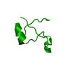 7dxtC 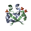 7dxuC  7dxvC 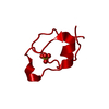 7dxwC  7dxxC 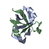 7dxyC 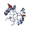 7dxzC  7dycC C: citing same article ( |
|---|---|
| Similar structure data |
-Links
 PDBj
PDBj
- Assembly
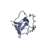
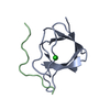
| Deposited unit | 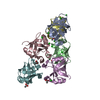
| ||||||||
|---|---|---|---|---|---|---|---|---|---|
| 1 | 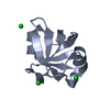
| ||||||||
| 2 | 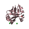
| ||||||||
| 3 | 
| ||||||||
| 4 | 
| ||||||||
| 5 | 
| ||||||||
| 6 | 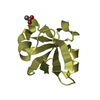
| ||||||||
| Unit cell |
|
-Components
| #1: Protein | Mass: 9353.974 Da / Num. of mol.: 6 Source method: isolated from a genetically manipulated source Source: (gene. exp.) synthetic construct (others) / Production host:  #2: Chemical | ChemComp-CL /   Mass: 35.453 Da / Num. of mol.: 9 / Source method: obtained synthetically / Formula: Cl Mass: 35.453 Da / Num. of mol.: 9 / Source method: obtained synthetically / Formula: Cl#3: Chemical | ChemComp-ACT /   Mass: 59.044 Da / Num. of mol.: 5 / Source method: obtained synthetically / Formula: C2H3O2 Mass: 59.044 Da / Num. of mol.: 5 / Source method: obtained synthetically / Formula: C2H3O2#4: Water | ChemComp-HOH / |  Mass: 18.015 Da / Num. of mol.: 716 / Source method: isolated from a natural source / Formula: H2O Mass: 18.015 Da / Num. of mol.: 716 / Source method: isolated from a natural source / Formula: H2OHas ligand of interest | N | |
|---|
-Experimental details
-Experiment
| Experiment | Method: X-RAY DIFFRACTION / Number of used crystals: 1 |
|---|
- Sample preparation
| Crystal | Density Matthews: 3.8 Å3/Da / Density % sol: 67.67 % |
|---|---|
| Crystal grow | Temperature: 293 K / Method: vapor diffusion, sitting drop Details: 100mM Sodium acetate pH3.8, 2M NaCl, 400 mM Lithium sulfate |
-Data collection
| Diffraction | Mean temperature: 100 K / Serial crystal experiment: N |
|---|---|
| Diffraction source | Source: SYNCHROTRON / Site: SPring-8 / Beamline: BL26B2 / Wavelength: 1 Å |
| Detector | Type: RAYONIX MX225-HS / Detector: CCD / Date: Apr 8, 2020 |
| Radiation | Protocol: SINGLE WAVELENGTH / Monochromatic (M) / Laue (L): M / Scattering type: x-ray |
| Radiation wavelength | Wavelength: 1 Å / Relative weight: 1 |
| Reflection | Resolution: 1.7→50 Å / Num. obs: 94555 / % possible obs: 99.6 % / Redundancy: 28 % / CC1/2: 1 / Net I/σ(I): 33 |
| Reflection shell | Resolution: 1.7→1.81 Å / Num. unique obs: 14747 / CC1/2: 0.96 |
- Processing
| Software |
| ||||||||||||||||||||||||||||||||||||||||||||||||||||||||||||||||||||||||||||||||||||||||||
|---|---|---|---|---|---|---|---|---|---|---|---|---|---|---|---|---|---|---|---|---|---|---|---|---|---|---|---|---|---|---|---|---|---|---|---|---|---|---|---|---|---|---|---|---|---|---|---|---|---|---|---|---|---|---|---|---|---|---|---|---|---|---|---|---|---|---|---|---|---|---|---|---|---|---|---|---|---|---|---|---|---|---|---|---|---|---|---|---|---|---|---|
| Refinement | Method to determine structure: MOLECULAR REPLACEMENT Starting model: Computationally Designed Mode Resolution: 1.705→47.298 Å / SU ML: 0.16 / Cross valid method: THROUGHOUT / σ(F): 1.49 / Phase error: 16.36 / Stereochemistry target values: ML
| ||||||||||||||||||||||||||||||||||||||||||||||||||||||||||||||||||||||||||||||||||||||||||
| Solvent computation | Shrinkage radii: 0.9 Å / VDW probe radii: 1.11 Å / Solvent model: FLAT BULK SOLVENT MODEL | ||||||||||||||||||||||||||||||||||||||||||||||||||||||||||||||||||||||||||||||||||||||||||
| Displacement parameters | Biso max: 110.86 Å2 / Biso mean: 30.9935 Å2 / Biso min: 12.56 Å2 | ||||||||||||||||||||||||||||||||||||||||||||||||||||||||||||||||||||||||||||||||||||||||||
| Refinement step | Cycle: final / Resolution: 1.705→47.298 Å
| ||||||||||||||||||||||||||||||||||||||||||||||||||||||||||||||||||||||||||||||||||||||||||
| LS refinement shell | Refine-ID: X-RAY DIFFRACTION / Rfactor Rfree error: 0
|
