 Movie
Movie Controller


+ Open data

- Basic information
| Entry | Database: PDB / ID: 1fd4 | ||||||
|---|---|---|---|---|---|---|---|
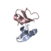
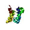
| Title | HUMAN BETA-DEFENSIN 2 | ||||||
 Components Components | BETA-DEFENSIN 2 | ||||||
 Keywords Keywords | ANTIMICROBIAL PEPTIDE / DEFENSIN / HUMAN BETA-DEFENSIN 2 / BETA-DEFENSIN | ||||||
| Function / homology |  Function and homology information Function and homology informationCCR6 chemokine receptor binding / Beta defensins / Defensins / antifungal innate immune response / chemoattractant activity / defense response to fungus / phosphatidylinositol-4,5-bisphosphate binding / cell chemotaxis / Golgi lumen / chemotaxis ...CCR6 chemokine receptor binding / Beta defensins / Defensins / antifungal innate immune response / chemoattractant activity / defense response to fungus / phosphatidylinositol-4,5-bisphosphate binding / cell chemotaxis / Golgi lumen / chemotaxis / antimicrobial humoral immune response mediated by antimicrobial peptide / killing of cells of another organism / defense response to Gram-negative bacterium / defense response to bacterium / defense response to Gram-positive bacterium / immune response / G protein-coupled receptor signaling pathway / : / extracellular region Similarity search - Function | ||||||
| Method |  X-RAY DIFFRACTION / SYNCHROTRON / MOLECULAR REPLACEMENT / Resolution: 1.7 Å X-RAY DIFFRACTION / SYNCHROTRON / MOLECULAR REPLACEMENT / Resolution: 1.7 Å | ||||||
 Authors Authors | Hoover, D.M. / Lubkowski, J. | ||||||
 Citation Citation | Journal: J.Biol.Chem. / Year: 2000 Title: The structure of human beta-defensin-2 shows evidence of higher order oligomerization. Authors: Hoover, D.M. / Rajashankar, K.R. / Blumenthal, R. / Puri, A. / Oppenheim, J.J. / Chertov, O. / Lubkowski, J. | ||||||
| History |
|
- Structure visualization
| Structure viewer | Molecule: MolmilJmol/JSmol |
|---|
- Downloads & links
-Download
| PDBx/mmCIF format | 1fd4.cif.gz | 150.4 KB | Display | PDBx/mmCIF format |
|---|---|---|---|---|
| PDB format | pdb1fd4.ent.gz | 120.6 KB | Display | PDB format |
| PDBx/mmJSON format | 1fd4.json.gz | Tree view | PDBx/mmJSON format | |
| Others |  Other downloads Other downloads |
-Validation report
| Arichive directory | https://data.pdbj.org/pub/pdb/validation_reports/fd/1fd4ftp://data.pdbj.org/pub/pdb/validation_reports/fd/1fd4 | HTTPS FTP |
|---|
-Related structure data


-Links
 PDBj
PDBj

- Assembly
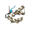
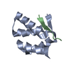
| Deposited unit | 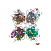
| ||||||||
|---|---|---|---|---|---|---|---|---|---|
| 1 | 
| ||||||||
| 2 | 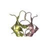
| ||||||||
| 3 | 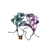
| ||||||||
| 4 | 
| ||||||||
| 5 | 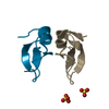
| ||||||||
| 6 | 
| ||||||||
| 7 | 
| ||||||||
| 8 | 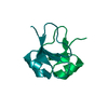
| ||||||||
| Unit cell |
| ||||||||
| Details | The biological assembly is a dimer. |
-Components
| #1: Protein/peptide | Mass: 4342.271 Da / Num. of mol.: 16 / Source method: obtained synthetically Details: THIS PEPTIDE WAS CHEMICALLY SYNTHESIZED. THE SEQUENCE OF THIS PEPTIDE OCCURS NATURALLY IN HUMANS (HOMO SAPIENS) References: UniProt: O15263 #2: Chemical | ChemComp-SO4 /   Mass: 96.063 Da / Num. of mol.: 5 / Source method: obtained synthetically / Formula: SO4 Mass: 96.063 Da / Num. of mol.: 5 / Source method: obtained synthetically / Formula: SO4#3: Water | ChemComp-HOH / |  Mass: 18.015 Da / Num. of mol.: 806 / Source method: isolated from a natural source / Formula: H2O Mass: 18.015 Da / Num. of mol.: 806 / Source method: isolated from a natural source / Formula: H2OHas protein modification | Y | |
|---|
-Experimental details
-Experiment
| Experiment | Method: X-RAY DIFFRACTION / Number of used crystals: 1 |
|---|
- Sample preparation
| Crystal | Density Matthews: 1 Å3/Da / Density % sol: 45.21 % | ||||||||||||||||||||||||
|---|---|---|---|---|---|---|---|---|---|---|---|---|---|---|---|---|---|---|---|---|---|---|---|---|---|
| Crystal grow | Temperature: 298.15 K / Method: vapor diffusion, hanging drop / pH: 8.5 Details: PEG 4000, tris, lithium sulfate, pH 8.5, VAPOR DIFFUSION, HANGING DROP, temperature 298.15K | ||||||||||||||||||||||||
| Crystal grow | *PLUS Details: used macroseeding | ||||||||||||||||||||||||
| Components of the solutions | *PLUS
|
-Data collection
| Diffraction | Mean temperature: 100 K |
|---|---|
| Diffraction source | Source: SYNCHROTRON / Site: NSLS  / Beamline: X9B / Wavelength: 0.979 / Beamline: X9B / Wavelength: 0.979 |
| Detector | Type: ADSC / Detector: CCD / Date: Oct 2, 1999 / Details: MIRRORS |
| Radiation | Monochromator: SI CRYSTAL / Protocol: SINGLE WAVELENGTH / Monochromatic (M) / Laue (L): M / Scattering type: x-ray |
| Radiation wavelength | Wavelength: 0.979 Å / Relative weight: 1 |
| Reflection | Resolution: 1.7→30 Å / Num. all: 65644 / Num. obs: 65644 / % possible obs: 96.3 % / Observed criterion σ(F): 0 / Observed criterion σ(I): 0 / Redundancy: 2.4 % / Biso Wilson estimate: 10.2 Å2 / Rmerge(I) obs: 0.043 / Net I/σ(I): 24.2 |
| Reflection shell | Resolution: 1.7→1.76 Å / Redundancy: 2.1 % / Rmerge(I) obs: 0.273 / Mean I/σ(I) obs: 4.7 / % possible all: 96.3 |
| Reflection | *PLUS % possible obs: 97.1 % / Num. measured all: 165583 |
| Reflection shell | *PLUS % possible obs: 96.3 % |
- Processing
| Software |
| |||||||||||||||||||||||||||||||||
|---|---|---|---|---|---|---|---|---|---|---|---|---|---|---|---|---|---|---|---|---|---|---|---|---|---|---|---|---|---|---|---|---|---|---|
| Refinement | Method to determine structure: MOLECULAR REPLACEMENT Starting model: HUMAN BETA-DEFESIN-2 Resolution: 1.7→30 Å / Num. parameters: 2327 / Num. restraintsaints: 2138 / Cross valid method: FREE R / σ(F): 0 / σ(I): 0 / Stereochemistry target values: ENGH AND HUBER
| |||||||||||||||||||||||||||||||||
| Refine analyze | Num. disordered residues: 3 / Occupancy sum hydrogen: 0 / Occupancy sum non hydrogen: 5615 | |||||||||||||||||||||||||||||||||
| Refinement step | Cycle: LAST / Resolution: 1.7→30 Å
| |||||||||||||||||||||||||||||||||
| Refine LS restraints |
| |||||||||||||||||||||||||||||||||
| Software | *PLUS Name: SHELXL-97 / Classification: refinement | |||||||||||||||||||||||||||||||||
| Refinement | *PLUS σ(F): 0 / % reflection Rfree: 11 % | |||||||||||||||||||||||||||||||||
| Solvent computation | *PLUS | |||||||||||||||||||||||||||||||||
| Displacement parameters | *PLUS | |||||||||||||||||||||||||||||||||
| Refine LS restraints | *PLUS
|
