Group: Data collection / Category: chem_comp_atom / chem_comp_bond
Remark 999
SEQUENCE P43M is the background mutation used in the Chazin lab to study all calbindin D9k mutants. ...SEQUENCE P43M is the background mutation used in the Chazin lab to study all calbindin D9k mutants. This mutation removes spectra-complicating cis-trans isomerization at Pro43 but does not otherwise affect the structure.
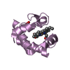
The full ensemble was ordered by lowest residual constraint violations, then the top 22 with favorable covalent geometries and AMBER energies were selected
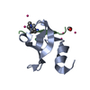
Representative
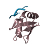
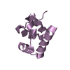
Model #1
closest to the average
-
Components
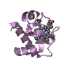
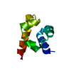
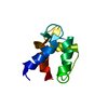
#1: Protein
calbindinD9k / CABP / Vitamin D-dependent calcium-binding protein / intestinal
Mass: 8454.515 Da / Num. of mol.: 1 Fragment: contains EF-HAND 1 (LOW AFFINITY) and EF-HAND 2 (HIGH AFFINITY) Mutation: F36G, P43M Source method: isolated from a genetically manipulated source Source: (gene. exp.) Bos taurus (domestic cattle) / Plasmid: pRCB1 / Species (production host): Escherichia coli / Production host: Escherichia coli BL21(DE3) (bacteria) / Strain (production host): BL21DE3 / References: UniProt: P02633
-
Experimental details
-
Experiment
Experiment
Method: SOLUTION NMR
NMR experiment
Conditions-ID
Experiment-ID
Solution-ID
Type
1
1
1
2D COSY
1
2
1
2D NOESY
1
3
1
2D TOCSY
1
4
3
2D NOESY
1
5
2
2D 15N-1H HSQC
1
6
2
3D 15N-separated TOCSY
1
7
2
3D 15N-separated NOESY
1
8
2
HNHA
1
9
2
HNHB
NMR details
Text: This structure was determined using a combination of standard 2D homonuclear and 15N-based 3D methods.
-
Sample preparation
Details
Solution-ID
Contents
Solvent system
1
2.5mMcalbindin; 95% H2O; 5% D2O
95% H20; 5% D2O
2
2.5 mM calbindin D9k U-15N; 95% H2O; 5% D2O
95% H2O/5% D2O
3
2.5mMcalbindinD9k; 100% D20
100% D2O
Sample conditions
Ionic strength: no added salts / pH: 6 / Pressure: ambient / Temperature: 300 K
Crystal grow
*PLUS
Method: other / Details: NMR
-
NMR measurement
Radiation
Protocol: SINGLE WAVELENGTH / Monochromatic (M) / Laue (L): M
Radiation wavelength
Relative weight: 1
NMR spectrometer
Type
Manufacturer
Model
Field strength (MHz)
Spectrometer-ID
Bruker AMX
Bruker
AMX
500
1
Bruker DMX
Bruker
DMX
750
2
-
Processing
NMR software
Name
Version
Developer
Classification
DIANA
2.8
Guntert
structuresolution
Amber
4.1
Pearlmann
structuresolution
Felix
97
MolecularSimulations, Inc.
dataanalysis
GLOMSA
unknown
Guntert
dataanalysis
GENXPK
1
Gippert
dataanalysis
Amber
4.1
Pearlmann
refinement
Refinement
Method: distance geometry, simulated annealing / Software ordinal: 1 Details: The structures are based on 1042 NOE restraints (186 intraresidue, 269 sequential, 289 medium range (2-4 residues apart), 298 long range), 18 hydrogen bond restraints (assigned as described ...Details: The structures are based on 1042 NOE restraints (186 intraresidue, 269 sequential, 289 medium range (2-4 residues apart), 298 long range), 18 hydrogen bond restraints (assigned as described in Skelton et al., 1995), and 115 dihedral constraints (45 phi, 42 psi, and 28 chi1).
NMR representative
Selection criteria: closest to the average
NMR ensemble
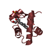
Conformer selection criteria: The full ensemble was ordered by lowest residual constraint violations, then the top 22 with favorable covalent geometries and AMBER energies were selected Conformers calculated total number: 50 / Conformers submitted total number: 22
+
About Yorodumi
-
News
-
Feb 9, 2022. New format data for meta-information of EMDB entries
New format data for meta-information of EMDB entries
Version 3 of the EMDB header file is now the official format.
The previous official version 1.9 will be removed from the archive.
In the structure databanks used in Yorodumi, some data are registered as the other names, "COVID-19 virus" and "2019-nCoV". Here are the details of the virus and the list of structure data.
Jan 31, 2019. EMDB accession codes are about to change! (news from PDBe EMDB page)
EMDB accession codes are about to change! (news from PDBe EMDB page)
The allocation of 4 digits for EMDB accession codes will soon come to an end. Whilst these codes will remain in use, new EMDB accession codes will include an additional digit and will expand incrementally as the available range of codes is exhausted. The current 4-digit format prefixed with “EMD-” (i.e. EMD-XXXX) will advance to a 5-digit format (i.e. EMD-XXXXX), and so on. It is currently estimated that the 4-digit codes will be depleted around Spring 2019, at which point the 5-digit format will come into force.
The EM Navigator/Yorodumi systems omit the EMD- prefix.
Related info.:Q: What is EMD? / ID/Accession-code notation in Yorodumi/EM Navigator
Yorodumi is a browser for structure data from EMDB, PDB, SASBDB, etc.
This page is also the successor to EM Navigator detail page, and also detail information page/front-end page for Omokage search.
The word "yorodu" (or yorozu) is an old Japanese word meaning "ten thousand". "mi" (miru) is to see.
Related info.:EMDB / PDB / SASBDB / Comparison of 3 databanks / Yorodumi Search / Aug 31, 2016. New EM Navigator & Yorodumi / Yorodumi Papers / Jmol/JSmol / Function and homology information / Changes in new EM Navigator and Yorodumi
 Movie
Movie Controller


 Open data
 Basic information
 Components
 Keywords
 Function and homology information
 Authors
 Citation
 Structure visualization
 Downloads & links
 Other downloads


 PDBj
PDBj Assembly
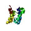

 HSQC
HSQC Sample preparation
 Processing