 Movie
Movie Controller

[English] 日本語
 Yorodumi
- PDB-7dty: Structural basis of ligand selectivity conferred by the human glu... -
+ Open data

- Basic information
| Entry | Database: PDB / ID: 7dty | ||||||||||||||||||||||||||||||||||||
|---|---|---|---|---|---|---|---|---|---|---|---|---|---|---|---|---|---|---|---|---|---|---|---|---|---|---|---|---|---|---|---|---|---|---|---|---|---|
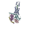
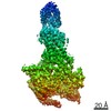
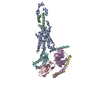
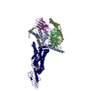
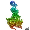
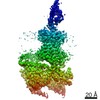
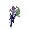
| Title | Structural basis of ligand selectivity conferred by the human glucose-dependent insulinotropic polypeptide receptor | ||||||||||||||||||||||||||||||||||||
 Components Components |
| ||||||||||||||||||||||||||||||||||||
 Keywords Keywords | STRUCTURAL PROTEIN / Glucose-dependent insulinotropic polypeptide receptor / cryo-electron microscopy / G protein-coupled receptor / ligand recognition | ||||||||||||||||||||||||||||||||||||
| Function / homology |  Function and homology information Function and homology informationgastric inhibitory polypeptide receptor binding / gastric inhibitory peptide signaling pathway / glucagon receptor binding / endocrine pancreas development / regulation of fatty acid biosynthetic process / Synthesis, secretion, and inactivation of Glucose-dependent Insulinotropic Polypeptide (GIP) / sensory perception of chemical stimulus / mu-type opioid receptor binding / corticotropin-releasing hormone receptor 1 binding / G-protein activation ...gastric inhibitory polypeptide receptor binding / gastric inhibitory peptide signaling pathway / glucagon receptor binding / endocrine pancreas development / regulation of fatty acid biosynthetic process / Synthesis, secretion, and inactivation of Glucose-dependent Insulinotropic Polypeptide (GIP) / sensory perception of chemical stimulus / mu-type opioid receptor binding / corticotropin-releasing hormone receptor 1 binding / G-protein activation / Activation of the phototransduction cascade / Glucagon-type ligand receptors / Thromboxane signalling through TP receptor / Sensory perception of sweet, bitter, and umami (glutamate) taste / G beta:gamma signalling through PI3Kgamma / G beta:gamma signalling through CDC42 / Cooperation of PDCL (PhLP1) and TRiC/CCT in G-protein beta folding / Activation of G protein gated Potassium channels / Inhibition of voltage gated Ca2+ channels via Gbeta/gamma subunits / Ca2+ pathway / G alpha (z) signalling events / High laminar flow shear stress activates signaling by PIEZO1 and PECAM1:CDH5:KDR in endothelial cells / Glucagon-like Peptide-1 (GLP1) regulates insulin secretion / Vasopressin regulates renal water homeostasis via Aquaporins / Adrenaline,noradrenaline inhibits insulin secretion / ADP signalling through P2Y purinoceptor 12 / G alpha (q) signalling events / beta-2 adrenergic receptor binding / G alpha (i) signalling events / Thrombin signalling through proteinase activated receptors (PARs) / Activation of G protein gated Potassium channels / G-protein activation / G beta:gamma signalling through PI3Kgamma / Prostacyclin signalling through prostacyclin receptor / G beta:gamma signalling through PLC beta / ADP signalling through P2Y purinoceptor 1 / Thromboxane signalling through TP receptor / Presynaptic function of Kainate receptors / G beta:gamma signalling through CDC42 / Inhibition of voltage gated Ca2+ channels via Gbeta/gamma subunits / G alpha (12/13) signalling events / Glucagon-type ligand receptors / G beta:gamma signalling through BTK / ADP signalling through P2Y purinoceptor 12 / Adrenaline,noradrenaline inhibits insulin secretion / Cooperation of PDCL (PhLP1) and TRiC/CCT in G-protein beta folding / Ca2+ pathway / G alpha (z) signalling events / Thrombin signalling through proteinase activated receptors (PARs) / Extra-nuclear estrogen signaling / G alpha (s) signalling events / G alpha (q) signalling events / response to starvation / photoreceptor outer segment membrane / spectrin binding / Glucagon-like Peptide-1 (GLP1) regulates insulin secretion / G alpha (i) signalling events / High laminar flow shear stress activates signaling by PIEZO1 and PECAM1:CDH5:KDR in endothelial cells / Vasopressin regulates renal water homeostasis via Aquaporins / exploration behavior / alkylglycerophosphoethanolamine phosphodiesterase activity / regulation of insulin secretion / photoreceptor outer segment / response to glucose / D1 dopamine receptor binding / adenylate cyclase-activating adrenergic receptor signaling pathway / adult locomotory behavior / insulin-like growth factor receptor binding / cardiac muscle cell apoptotic process / photoreceptor inner segment / ionotropic glutamate receptor binding / sensory perception of pain / adenylate cyclase activator activity / hormone activity / memory / G-protein beta/gamma-subunit complex binding / positive regulation of insulin secretion / Glucagon-type ligand receptors / cellular response to catecholamine stimulus / adenylate cyclase-activating dopamine receptor signaling pathway / cellular response to prostaglandin E stimulus / heterotrimeric G-protein complex / G-protein beta-subunit binding / positive regulation of cytosolic calcium ion concentration / sensory perception of taste / signaling receptor complex adaptor activity / adenylate cyclase-activating G protein-coupled receptor signaling pathway / retina development in camera-type eye / cell body / GTPase binding / secretory granule lumen / cellular response to hypoxia / G alpha (s) signalling events / phospholipase C-activating G protein-coupled receptor signaling pathway / Hydrolases; Acting on acid anhydrides; Acting on GTP to facilitate cellular and subcellular movement / cell population proliferation / endoplasmic reticulum lumen / G protein-coupled receptor signaling pathway / GTPase activity / dendrite Similarity search - Function | ||||||||||||||||||||||||||||||||||||
| Biological species |  Homo sapiens (human) Homo sapiens (human) synthetic construct (others) | ||||||||||||||||||||||||||||||||||||
| Method | ELECTRON MICROSCOPY / single particle reconstruction / cryo EM / Resolution: 2.98 Å | ||||||||||||||||||||||||||||||||||||
 Authors Authors | Zhao, F.H. / Zhang, C. / Zhou, Q.T. / Hang, K.N. / Zou, X.Y. / Chen, Y. / Wu, F. / Rao, Q.D. / Dai, A.T. / Yin, W.C. ...Zhao, F.H. / Zhang, C. / Zhou, Q.T. / Hang, K.N. / Zou, X.Y. / Chen, Y. / Wu, F. / Rao, Q.D. / Dai, A.T. / Yin, W.C. / Shen, D.D. / Zhang, Y. / Xia, T. / Stevens, R.C. / Xu, H.E. / Yang, D.H. / Zhao, L.H. / Wang, M.W. | ||||||||||||||||||||||||||||||||||||
| Funding support |  China, 4items China, 4items
| ||||||||||||||||||||||||||||||||||||
 Citation Citation | Journal: Elife / Year: 2021 Title: Structural insights into hormone recognition by the human glucose-dependent insulinotropic polypeptide receptor. Authors: Fenghui Zhao / Chao Zhang / Qingtong Zhou / Kaini Hang / Xinyu Zou / Yan Chen / Fan Wu / Qidi Rao / Antao Dai / Wanchao Yin / Dan-Dan Shen / Yan Zhang / Tian Xia / Raymond C Stevens / H Eric ...Authors: Fenghui Zhao / Chao Zhang / Qingtong Zhou / Kaini Hang / Xinyu Zou / Yan Chen / Fan Wu / Qidi Rao / Antao Dai / Wanchao Yin / Dan-Dan Shen / Yan Zhang / Tian Xia / Raymond C Stevens / H Eric Xu / Dehua Yang / Lihua Zhao / Ming-Wei Wang / Abstract: Glucose-dependent insulinotropic polypeptide (GIP) is a peptide hormone that exerts crucial metabolic functions by binding and activating its cognate receptor, GIPR. As an important therapeutic ...Glucose-dependent insulinotropic polypeptide (GIP) is a peptide hormone that exerts crucial metabolic functions by binding and activating its cognate receptor, GIPR. As an important therapeutic target, GIPR has been subjected to intensive structural studies without success. Here, we report the cryo-EM structure of the human GIPR in complex with GIP and a G heterotrimer at a global resolution of 2.9 Å. GIP adopts a single straight helix with its N terminus dipped into the receptor transmembrane domain (TMD), while the C terminus is closely associated with the extracellular domain and extracellular loop 1. GIPR employs conserved residues in the lower half of the TMD pocket to recognize the common segments shared by GIP homologous peptides, while uses non-conserved residues in the upper half of the TMD pocket to interact with residues specific for GIP. These results provide a structural framework of hormone recognition and GIPR activation. | ||||||||||||||||||||||||||||||||||||
| History |
|
- Structure visualization
| Movie |
Movie viewer |
|---|---|
| Structure viewer | Molecule: MolmilJmol/JSmol |
 UCSF Chimera
- Downloads & links
-Download
| PDBx/mmCIF format | 7dty.cif.gz | 230.3 KB | Display | PDBx/mmCIF format |
|---|---|---|---|---|
| PDB format | pdb7dty.ent.gz | 176.6 KB | Display | PDB format |
| PDBx/mmJSON format | 7dty.json.gz | Tree view | PDBx/mmJSON format | |
| Others |  Other downloads Other downloads |
-Validation report
| Arichive directory | https://data.pdbj.org/pub/pdb/validation_reports/dt/7dtyftp://data.pdbj.org/pub/pdb/validation_reports/dt/7dty | HTTPS FTP |
|---|
-Related structure data
| Related structure data |  30860MC M: map data used to model this data C: citing same article ( |
|---|---|
| Similar structure data |


-Links
 PDBj
PDBj


- Assembly
| Deposited unit | 
|
|---|---|
| 1 |
|
-Components
-Guanine nucleotide-binding protein ... , 3 types, 3 molecules ABG
| #3: Protein | Mass: 45727.441 Da / Num. of mol.: 1 Source method: isolated from a genetically manipulated source Source: (gene. exp.)   Spodoptera frugiperda (fall armyworm) / References: UniProt: P04896 Spodoptera frugiperda (fall armyworm) / References: UniProt: P04896 |
|---|---|
| #4: Protein | Mass: 40226.992 Da / Num. of mol.: 1 Source method: isolated from a genetically manipulated source Source: (gene. exp.) Spodoptera frugiperda (fall armyworm) / References: UniProt: P54311 |
| #5: Protein | Mass: 7861.143 Da / Num. of mol.: 1 Source method: isolated from a genetically manipulated source Source: (gene. exp.) Spodoptera frugiperda (fall armyworm) / References: UniProt: P63212 |
-Protein / Protein/peptide / Antibody / Non-polymers , 4 types, 9 molecules RPN
| #1: Protein | Mass: 64950.695 Da / Num. of mol.: 1 Source method: isolated from a genetically manipulated source Details: mutations:1 / Source: (gene. exp.) Homo sapiens (human) / Production host: Spodoptera frugiperda (fall armyworm) |
|---|---|
| #2: Protein/peptide | Mass: 4990.586 Da / Num. of mol.: 1 / Source method: obtained synthetically / Source: (synth.) Homo sapiens (human) / References: UniProt: P09681 |
| #6: Antibody | Mass: 15343.019 Da / Num. of mol.: 1 Source method: isolated from a genetically manipulated source Source: (gene. exp.) synthetic construct (others) / Production host:  |
| #7: Chemical | ChemComp-CLR /  Mass: 386.654 Da / Num. of mol.: 6 / Source method: obtained synthetically / Formula: C27H46O / Feature type: SUBJECT OF INVESTIGATION Mass: 386.654 Da / Num. of mol.: 6 / Source method: obtained synthetically / Formula: C27H46O / Feature type: SUBJECT OF INVESTIGATION |
-Details
| Has ligand of interest | Y |
|---|---|
| Has protein modification | Y |
-Experimental details
-Experiment
| Experiment | Method: ELECTRON MICROSCOPY |
|---|---|
| EM experiment | Aggregation state: PARTICLE / 3D reconstruction method: single particle reconstruction |
- Sample preparation
| Component | Name: Cryo-EM structure of the human glucose-dependent insulinotropic polypeptide receptor in complex with GIP and G protein Type: COMPLEX / Entity ID: #1-#6 / Source: RECOMBINANT |
|---|---|
| Source (natural) | Organism: Homo sapiens (human) |
| Source (recombinant) | Organism: Spodoptera frugiperda (fall armyworm) |
| Buffer solution | pH: 7.4 |
| Specimen | Embedding applied: NO / Shadowing applied: NO / Staining applied: NO / Vitrification applied: YES |
| Vitrification | Cryogen name: ETHANE |
- Electron microscopy imaging
| Experimental equipment |  Model: Titan Krios / Image courtesy: FEI Company |
|---|---|
| Microscopy | Model: FEI TITAN KRIOS |
| Electron gun | Electron source: OTHER / Accelerating voltage: 300 kV / Illumination mode: OTHER |
| Electron lens | Mode: BRIGHT FIELD |
| Image recording | Electron dose: 80 e/Å2 / Film or detector model: GATAN K3 (6k x 4k) |
- Processing
| Software | Name: PHENIX / Version: 1.13_2998: / Classification: refinement |
|---|---|
| EM software | Name: PHENIX / Category: model refinement |
| CTF correction | Type: PHASE FLIPPING AND AMPLITUDE CORRECTION |
| 3D reconstruction | Resolution: 2.98 Å / Resolution method: DIFFRACTION PATTERN/LAYERLINES / Num. of particles: 295021 / Symmetry type: POINT |
