 Movie
Movie Controller

[English] 日本語
 Yorodumi
- EMDB-30048: CryoEM structure of human PAC1 receptor in complex with PACAP38 -
+ Open data

- Basic information
| Entry | Database: EMDB / ID: EMD-30048 | |||||||||
|---|---|---|---|---|---|---|---|---|---|---|
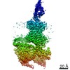
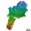
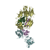
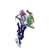
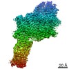
| Title | CryoEM structure of human PAC1 receptor in complex with PACAP38 | |||||||||
 Map data Map data | ||||||||||
 Sample Sample |
| |||||||||
 Keywords Keywords | GPCR / PROTEIN BINDING | |||||||||
| Function / homology |  Function and homology information Function and homology informationnegative regulation of response to reactive oxygen species / pituitary adenylate cyclase activating polypeptide activity / development of primary female sexual characteristics / type 1 vasoactive intestinal polypeptide receptor binding / type 2 vasoactive intestinal polypeptide receptor binding / pituitary adenylate cyclase-activating polypeptide receptor activity / vasoactive intestinal polypeptide receptor activity / positive regulation of growth hormone secretion / positive regulation of chemokine (C-C motif) ligand 5 production / positive regulation of small GTPase mediated signal transduction ...negative regulation of response to reactive oxygen species / pituitary adenylate cyclase activating polypeptide activity / development of primary female sexual characteristics / type 1 vasoactive intestinal polypeptide receptor binding / type 2 vasoactive intestinal polypeptide receptor binding / pituitary adenylate cyclase-activating polypeptide receptor activity / vasoactive intestinal polypeptide receptor activity / positive regulation of growth hormone secretion / positive regulation of chemokine (C-C motif) ligand 5 production / positive regulation of small GTPase mediated signal transduction / neuropeptide hormone activity / regulation of G protein-coupled receptor signaling pathway / NGF-independant TRKA activation / G protein-coupled peptide receptor activity / positive regulation of protein kinase activity / neuropeptide binding / insulin secretion / peptide hormone receptor binding / positive regulation of inositol phosphate biosynthetic process / adenylate cyclase-activating G protein-coupled bile acid receptor signaling pathway / adenylate cyclase-activating serotonin receptor signaling pathway / negative regulation of cell cycle / positive regulation of cAMP/PKA signal transduction / peptide hormone binding / regulation of skeletal muscle contraction / adenylate cyclase binding / positive regulation of GTPase activity / PKA activation in glucagon signalling / positive regulation of calcium ion transport into cytosol / hair follicle placode formation / bicellular tight junction / developmental growth / intracellular transport / cAMP/PKA signal transduction / multicellular organismal response to stress / D1 dopamine receptor binding / vascular endothelial cell response to laminar fluid shear stress / renal water homeostasis / activation of adenylate cyclase activity / Hedgehog 'off' state / adenylate cyclase-activating adrenergic receptor signaling pathway / cellular response to acidic pH / cellular response to glucagon stimulus / intracellular glucose homeostasis / adenylate cyclase activator activity / positive regulation of insulin secretion involved in cellular response to glucose stimulus / trans-Golgi network membrane / negative regulation of inflammatory response to antigenic stimulus / female pregnancy / neuropeptide signaling pathway / response to prostaglandin E / bone development / caveola / platelet aggregation / cognition / small GTPase binding / G-protein beta/gamma-subunit complex binding / adenylate cyclase-modulating G protein-coupled receptor signaling pathway / neuron projection development / positive regulation of insulin secretion / Olfactory Signaling Pathway / Activation of the phototransduction cascade / G protein-coupled acetylcholine receptor signaling pathway / G beta:gamma signalling through PLC beta / Presynaptic function of Kainate receptors / Thromboxane signalling through TP receptor / Activation of G protein gated Potassium channels / Inhibition of voltage gated Ca2+ channels via Gbeta/gamma subunits / G-protein activation / sensory perception of smell / Glucagon signaling in metabolic regulation / Prostacyclin signalling through prostacyclin receptor / G beta:gamma signalling through CDC42 / Synthesis, secretion, and inactivation of Glucagon-like Peptide-1 (GLP-1) / G beta:gamma signalling through BTK / photoreceptor disc membrane / ADP signalling through P2Y purinoceptor 12 / response to estradiol / Glucagon-type ligand receptors / Sensory perception of sweet, bitter, and umami (glutamate) taste / Adrenaline,noradrenaline inhibits insulin secretion / Vasopressin regulates renal water homeostasis via Aquaporins / Glucagon-like Peptide-1 (GLP1) regulates insulin secretion / G alpha (z) signalling events / cellular response to catecholamine stimulus / ADP signalling through P2Y purinoceptor 1 / ADORA2B mediated anti-inflammatory cytokines production / G beta:gamma signalling through PI3Kgamma / cell-cell signaling / adenylate cyclase-activating dopamine receptor signaling pathway / regulation of protein localization / Cooperation of PDCL (PhLP1) and TRiC/CCT in G-protein beta folding / positive regulation of cold-induced thermogenesis / GPER1 signaling / cellular response to prostaglandin E stimulus / heterotrimeric G-protein complex / G alpha (12/13) signalling events / G-protein beta-subunit binding / Inactivation, recovery and regulation of the phototransduction cascade / extracellular vesicle Similarity search - Function | |||||||||
| Biological species |  Homo sapiens (human) / Homo sapiens (human) /  | |||||||||
| Method | single particle reconstruction / cryo EM / Resolution: 3.5 Å | |||||||||
 Authors Authors | Song X / Wang J | |||||||||
 Citation Citation | Journal: Cell Res / Year: 2020 Title: Cryo-EM structures of PAC1 receptor reveal ligand binding mechanism. Authors: Jia Wang / Xianqiang Song / Dandan Zhang / Xiaoqing Chen / Xun Li / Yaping Sun / Cui Li / Yunpeng Song / Yao Ding / Ruobing Ren / Essa Hu Harrington / Liaoyuan A Hu / Wenge Zhong / Cen Xu / ...Authors: Jia Wang / Xianqiang Song / Dandan Zhang / Xiaoqing Chen / Xun Li / Yaping Sun / Cui Li / Yunpeng Song / Yao Ding / Ruobing Ren / Essa Hu Harrington / Liaoyuan A Hu / Wenge Zhong / Cen Xu / Xin Huang / Hong-Wei Wang / Yingli Ma /   Abstract: The pituitary adenylate cyclase-activating polypeptide type I receptor (PAC1R) belongs to the secretin receptor family and is widely distributed in the central neural system and peripheral organs. ...The pituitary adenylate cyclase-activating polypeptide type I receptor (PAC1R) belongs to the secretin receptor family and is widely distributed in the central neural system and peripheral organs. Abnormal activation of the receptor mediates trigeminovascular activation and sensitization, which is highly related to migraine, making PAC1R a potential therapeutic target. Elucidation of PAC1R activation mechanism would benefit discovery of therapeutic drugs for neuronal disorders. PAC1R activity is governed by pituitary adenylate cyclase-activating polypeptide (PACAP), known as a major vasodilator neuropeptide, and maxadilan, a native peptide from the sand fly, which is also capable of activating the receptor with similar potency. These peptide ligands have divergent sequences yet initiate convergent PAC1R activity. It is of interest to understand the mechanism of PAC1R ligand recognition and receptor activity regulation through structural biology. Here we report two near-atomic resolution cryo-EM structures of PAC1R activated by PACAP38 or maxadilan, providing structural insights into two distinct ligand binding modes. The structures illustrate flexibility of the extracellular domain (ECD) for ligands with distinct conformations, where ECD accommodates ligands in different orientations while extracellular loop 1 (ECL1) protrudes to further anchor the ligand bound in the orthosteric site. By structure-guided molecular modeling and mutagenesis, we tested residues in the ligand-binding pockets and identified clusters of residues that are critical for receptor activity. The structures reported here for the first time elucidate the mechanism of specificity and flexibility of ligand recognition and binding for PAC1R, and provide insights toward the design of therapeutic molecules targeting PAC1R. | |||||||||
| History |
|
- Structure visualization
| Movie |
Movie viewer |
|---|---|
| Structure viewer | EM map: SurfViewMolmilJmol/JSmol |
| Supplemental images |

- Downloads & links
-EMDB archive
| Map data | emd_30048.map.gz | 2.5 MB | EMDB map data format | |
|---|---|---|---|---|
| Header (meta data) | emd-30048-v30.xmlemd-30048.xml | 16.9 KB 16.9 KB | Display Display | EMDB header |
| Images | 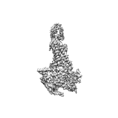 emd_30048.png emd_30048.png | 56.7 KB | ||
| Filedesc metadata | emd-30048.cif.gz | 6.5 KB | ||
| Archive directory |  http://ftp.pdbj.org/pub/emdb/structures/EMD-30048ftp://ftp.pdbj.org/pub/emdb/structures/EMD-30048 http://ftp.pdbj.org/pub/emdb/structures/EMD-30048ftp://ftp.pdbj.org/pub/emdb/structures/EMD-30048 | HTTPS FTP |
-Related structure data
| Related structure data |  6m1iMC  6m1hC C: citing same article ( M: atomic model generated by this map |
|---|---|
| Similar structure data |


-Links
| EMDB pages | EMDB (EBI/PDBe) / EMDataResource |
|---|---|
| Related items in Molecule of the Month |


-Map
| File | Download / File: emd_30048.map.gz / Format: CCP4 / Size: 22.2 MB / Type: IMAGE STORED AS FLOATING POINT NUMBER (4 BYTES) | ||||||||||||||||||||||||||||||||||||||||||||||||||||||||||||||||||||
|---|---|---|---|---|---|---|---|---|---|---|---|---|---|---|---|---|---|---|---|---|---|---|---|---|---|---|---|---|---|---|---|---|---|---|---|---|---|---|---|---|---|---|---|---|---|---|---|---|---|---|---|---|---|---|---|---|---|---|---|---|---|---|---|---|---|---|---|---|---|
| Projections & slices | Image control
Images are generated by Spider. | ||||||||||||||||||||||||||||||||||||||||||||||||||||||||||||||||||||
| Voxel size | X=Y=Z: 1.091 Å | ||||||||||||||||||||||||||||||||||||||||||||||||||||||||||||||||||||
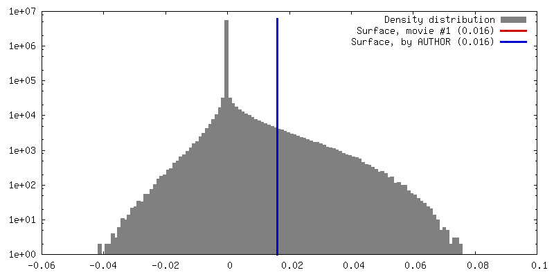
| Density |
| ||||||||||||||||||||||||||||||||||||||||||||||||||||||||||||||||||||
| Symmetry | Space group: 1 | ||||||||||||||||||||||||||||||||||||||||||||||||||||||||||||||||||||
| Details | EMDB XML:
CCP4 map header:
| ||||||||||||||||||||||||||||||||||||||||||||||||||||||||||||||||||||
 Z (Sec.)
 Y (Row.)
 X (Col.)


-Supplemental data
- Sample components
-Entire : PACAP38-PAC1R complex
| Entire | Name: PACAP38-PAC1R complex |
|---|---|
| Components |
|
-Supramolecule #1: PACAP38-PAC1R complex
| Supramolecule | Name: PACAP38-PAC1R complex / type: complex / ID: 1 / Parent: 0 / Macromolecule list: all |
|---|---|
| Source (natural) | Organism: Homo sapiens (human) |
| Molecular weight | Theoretical: 150 KDa |
-Macromolecule #1: Pituitary adenylate cyclase-activating polypeptide type I receptor
| Macromolecule | Name: Pituitary adenylate cyclase-activating polypeptide type I receptor type: protein_or_peptide / ID: 1 / Number of copies: 1 / Enantiomer: LEVO |
|---|---|
| Source (natural) | Organism: Homo sapiens (human) |
| Molecular weight | Theoretical: 47.315629 KDa |
| Recombinant expression | Organism:  Insect BA phytoplasma (bacteria) Insect BA phytoplasma (bacteria) |
| Sequence | String: DYKDDDDKIF KKEQAMCLEK IQRANELMGF NDSSPGCPGM WDNITCWKPA HVGEMVLVSC PELFRIFNPD QDMGVVSRNC TEDGWSEPF PHYFDACGFD EYESETGDQD YYYLSVKALY TVGYSLSLVA LLLAMVILCR FRKLHCTRNF IHMNLFVSFM L RAISVFIK ...String: DYKDDDDKIF KKEQAMCLEK IQRANELMGF NDSSPGCPGM WDNITCWKPA HVGEMVLVSC PELFRIFNPD QDMGVVSRNC TEDGWSEPF PHYFDACGFD EYESETGDQD YYYLSVKALY TVGYSLSLVA LLLAMVILCR FRKLHCTRNF IHMNLFVSFM L RAISVFIK DWILYAEQDS NHCFISTVEC KAVMVFFHYC VVSNYFWLFI EGLYLFTLLV ETFFPERRYF YWYTIIGWGA PL VFVTVWA TLRLYFDDTG CWDMNDSTAL WWVIKGPVVG SIMVNFVLFI GIIVILVQKL QSPDMGGNES SIYLRLARST LLL IPLFGI HYTVFAFSPE NVSKRERLVF ELGLGSFQGF VVAVLYCFLN GEVQAEIKRK WRSWKVNRYF AVDFKHRHPS LASS LEVLF Q |
-Macromolecule #2: Pituitary adenylate cyclase-activating polypeptide
| Macromolecule | Name: Pituitary adenylate cyclase-activating polypeptide / type: protein_or_peptide / ID: 2 / Number of copies: 1 / Enantiomer: LEVO |
|---|---|
| Source (natural) | Organism: Homo sapiens (human) |
| Molecular weight | Theoretical: 4.547336 KDa |
| Sequence | String: HSDGIFTDSY SRYRKQMAVK KYLAAVLGKR YKQRVKNK UniProtKB: Pituitary adenylate cyclase-activating polypeptide |
-Macromolecule #3: Nanobody 35
| Macromolecule | Name: Nanobody 35 / type: protein_or_peptide / ID: 3 / Number of copies: 1 / Enantiomer: LEVO |
|---|---|
| Source (natural) | Organism: |
| Molecular weight | Theoretical: 14.71432 KDa |
| Recombinant expression | Organism: |
| Sequence | String: QVQLQESGGG LVQPGGSLRL SCAASGFTFS NYKMNWVRQA PGKGLEWVSD ISQSGASISY TGSVKGRFTI SRDNAKNTLY LQMNSLKPE DTAVYYCARC PAPFTRDCFD VTSTTYAYRG QGTQVTVSSH HHHHH |
-Macromolecule #4: Guanine nucleotide-binding protein G(I)/G(S)/G(O) subunit gamma-2
| Macromolecule | Name: Guanine nucleotide-binding protein G(I)/G(S)/G(O) subunit gamma-2 type: protein_or_peptide / ID: 4 / Number of copies: 1 / Enantiomer: LEVO |
|---|---|
| Source (natural) | Organism: Homo sapiens (human) |
| Molecular weight | Theoretical: 7.861143 KDa |
| Recombinant expression | Organism: Insect BA phytoplasma (bacteria) |
| Sequence | String: MASNNTASIA QARKLVEQLK MEANIDRIKV SKAAADLMAY CEAHAKEDPL LTPVPASENP FREKKFFCAI L UniProtKB: Guanine nucleotide-binding protein G(I)/G(S)/G(O) subunit gamma-2 |
-Macromolecule #5: Guanine nucleotide-binding protein G(I)/G(S)/G(T) subunit beta-1
| Macromolecule | Name: Guanine nucleotide-binding protein G(I)/G(S)/G(T) subunit beta-1 type: protein_or_peptide / ID: 5 / Number of copies: 1 / Enantiomer: LEVO |
|---|---|
| Source (natural) | Organism: Homo sapiens (human) |
| Molecular weight | Theoretical: 37.47398 KDa |
| Recombinant expression | Organism: Insect BA phytoplasma (bacteria) |
| Sequence | String: GMSELDQLRQ EAEQLKNQIR DARKACADAT LSQITNNIDP VGRIQMRTRR TLRGHLAKIY AMHWGTDSRL LVSASQDGKL IIWDSYTTN KVHAIPLRSS WVMTCAYAPS GNYVACGGLD NICSIYNLKT REGNVRVSRE LAGHTGYLSC CRFLDDNQIV T SSGDTTCA ...String: GMSELDQLRQ EAEQLKNQIR DARKACADAT LSQITNNIDP VGRIQMRTRR TLRGHLAKIY AMHWGTDSRL LVSASQDGKL IIWDSYTTN KVHAIPLRSS WVMTCAYAPS GNYVACGGLD NICSIYNLKT REGNVRVSRE LAGHTGYLSC CRFLDDNQIV T SSGDTTCA LWDIETGQQT TTFTGHTGDV MSLSLAPDTR LFVSGACDAS AKLWDVREGM CRQTFTGHES DINAICFFPN GN AFATGSD DATCRLFDLR ADQELMTYSH DNIICGITSV SFSKSGRLLL AGYDDFNCNV WDALKADRAG VLAGHDNRVS CLG VTDDGM AVATGSWDSF LKIWN UniProtKB: Guanine nucleotide-binding protein G(I)/G(S)/G(T) subunit beta-1 |
-Macromolecule #6: Guanine nucleotide-binding protein G(s) subunit alpha isoforms short
| Macromolecule | Name: Guanine nucleotide-binding protein G(s) subunit alpha isoforms short type: protein_or_peptide / ID: 6 / Number of copies: 1 / Enantiomer: LEVO |
|---|---|
| Source (natural) | Organism: Homo sapiens (human) |
| Molecular weight | Theoretical: 45.683434 KDa |
| Recombinant expression | Organism: Insect BA phytoplasma (bacteria) |
| Sequence | String: MGCLGNSKTE DQRNEEKAQR EANKKIEKQL QKDKQVYRAT HRLLLLGAGE SGKNTIVKQM RILHVNGFNG EGGEEDPQAA RSNSDGEKA TKVQDIKNNL KEAIETIVAA MSNLVPPVEL ANPENQFRVD YILSVMNVPD FDFPPEFYEH AKALWEDEGV R ACYERSNE ...String: MGCLGNSKTE DQRNEEKAQR EANKKIEKQL QKDKQVYRAT HRLLLLGAGE SGKNTIVKQM RILHVNGFNG EGGEEDPQAA RSNSDGEKA TKVQDIKNNL KEAIETIVAA MSNLVPPVEL ANPENQFRVD YILSVMNVPD FDFPPEFYEH AKALWEDEGV R ACYERSNE YQLIDCAQYF LDKIDVIKQA DYVPSDQDLL RCRVLTSGIF ETKFQVDKVN FHMFDVGAQR DERRKWIQCF ND VTAIIFV VASSSYNMVI REDNQTNRLQ AALKLFDSIW NNKWLRDTSV ILFLNKQDLL AEKVLAGKSK IEDYFPEFAR YTT PEDATP EPGEDPRVTR AKYFIRDEFL RISTASGDGR HYCYPHFTCA VDTENIRRVF NDCRDIIQRM HLRQYELL |
-Experimental details
-Structure determination
| Method | cryo EM |
|---|---|
 Processing Processing | single particle reconstruction |
| Aggregation state | particle |
-Sample preparation
| Concentration | 5 mg/mL |
|---|---|
| Buffer | pH: 7.5 |
| Vitrification | Cryogen name: ETHANE / Chamber humidity: 100 % / Instrument: FEI VITROBOT MARK IV |
- Electron microscopy
| Microscope | FEI TITAN KRIOS |
|---|---|
| Image recording | Film or detector model: GATAN K2 SUMMIT (4k x 4k) / Detector mode: SUPER-RESOLUTION / Digitization - Dimensions - Width: 3838 pixel / Digitization - Dimensions - Height: 3710 pixel / Average electron dose: 50.0 e/Å2 |
| Electron beam | Acceleration voltage: 300 kV / Electron source:  FIELD EMISSION GUN FIELD EMISSION GUN |
| Electron optics | C2 aperture diameter: 50.0 µm / Illumination mode: FLOOD BEAM / Imaging mode: BRIGHT FIELD / Cs: 0.0 mm |
| Experimental equipment |  Model: Titan Krios / Image courtesy: FEI Company |
-Image processing
| Startup model | Type of model: OTHER |
|---|---|
| Final reconstruction | Number classes used: 1 / Applied symmetry - Point group: C1 (asymmetric) / Resolution.type: BY AUTHOR / Resolution: 3.5 Å / Resolution method: FSC 0.143 CUT-OFF / Software - Name: RELION (ver. 3.0) / Number images used: 82970 |
| Initial angle assignment | Type: MAXIMUM LIKELIHOOD |
| Final angle assignment | Type: MAXIMUM LIKELIHOOD |

