 Movie
Movie Controller

[English] 日本語
 Yorodumi
- PDB-7d8l: The structure of nucleoside phosphatase Sa1684 complex with GTP a... -
+ Open data

- Basic information
| Entry | Database: PDB / ID: 7d8l | ||||||
|---|---|---|---|---|---|---|---|
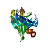
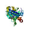
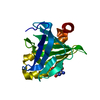
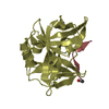
| Title | The structure of nucleoside phosphatase Sa1684 complex with GTP analogue from Staphylococcus aureus | ||||||
 Components Components | UPF0374 protein SA1684 | ||||||
 Keywords Keywords | CYTOSOLIC PROTEIN / nucleotide phosphatase | ||||||
| Function / homology |  Function and homology information Function and homology informationnucleoside diphosphate phosphatase / nucleoside diphosphate phosphatase activity / ribonucleoside triphosphate phosphatase activity / nucleoside-triphosphate phosphatase / magnesium ion binding Similarity search - Function | ||||||
| Biological species |  Staphylococcus aureus subsp. aureus N315 (bacteria) Staphylococcus aureus subsp. aureus N315 (bacteria) | ||||||
| Method |  X-RAY DIFFRACTION / SYNCHROTRON / SAD / Resolution: 1.55 Å X-RAY DIFFRACTION / SYNCHROTRON / SAD / Resolution: 1.55 Å | ||||||
 Authors Authors | Wang, Z. / Li, X. | ||||||
| Funding support |  China, 1items China, 1items
| ||||||
 Citation Citation | Journal: Febs J. / Year: 2021 Title: The structural mechanism for the nucleoside tri- and diphosphate hydrolysis activity of Ntdp from Staphylococcus aureus. Authors: Wang, Z. / Shen, H. / He, B. / Teng, M. / Guo, Q. / Li, X. | ||||||
| History |
|
- Structure visualization
| Structure viewer | Molecule: MolmilJmol/JSmol |
|---|
- Downloads & links
-Download
| PDBx/mmCIF format | 7d8l.cif.gz | 56.4 KB | Display | PDBx/mmCIF format |
|---|---|---|---|---|
| PDB format | pdb7d8l.ent.gz | 38.7 KB | Display | PDB format |
| PDBx/mmJSON format | 7d8l.json.gz | Tree view | PDBx/mmJSON format | |
| Others |  Other downloads Other downloads |
-Validation report
| Arichive directory | https://data.pdbj.org/pub/pdb/validation_reports/d8/7d8lftp://data.pdbj.org/pub/pdb/validation_reports/d8/7d8l | HTTPS FTP |
|---|
-Related structure data


-Links
 PDBj
PDBj

- Assembly
| Deposited unit | 
| ||||||||
|---|---|---|---|---|---|---|---|---|---|
| 1 |
| ||||||||
| Unit cell |
|
-Components
| #1: Protein | Mass: 21734.592 Da / Num. of mol.: 1 Source method: isolated from a genetically manipulated source Source: (gene. exp.) Staphylococcus aureus subsp. aureus N315 (bacteria)Strain: N315 / Gene: SA1684 / Production host: | ||||||
|---|---|---|---|---|---|---|---|
| #2: Chemical | ChemComp-GSP /   Mass: 539.246 Da / Num. of mol.: 1 / Source method: obtained synthetically / Formula: C10H16N5O13P3S / Feature type: SUBJECT OF INVESTIGATION Mass: 539.246 Da / Num. of mol.: 1 / Source method: obtained synthetically / Formula: C10H16N5O13P3S / Feature type: SUBJECT OF INVESTIGATION | ||||||
| #3: Chemical |   Mass: 24.305 Da / Num. of mol.: 2 / Source method: obtained synthetically / Formula: Mg / Feature type: SUBJECT OF INVESTIGATION Mass: 24.305 Da / Num. of mol.: 2 / Source method: obtained synthetically / Formula: Mg / Feature type: SUBJECT OF INVESTIGATION#4: Chemical | ChemComp-GOL / |   Mass: 92.094 Da / Num. of mol.: 1 / Source method: obtained synthetically / Formula: C3H8O3 Mass: 92.094 Da / Num. of mol.: 1 / Source method: obtained synthetically / Formula: C3H8O3#5: Water | ChemComp-HOH / |  Mass: 18.015 Da / Num. of mol.: 133 / Source method: isolated from a natural source / Formula: H2O Mass: 18.015 Da / Num. of mol.: 133 / Source method: isolated from a natural source / Formula: H2OHas ligand of interest | Y | |
-Experimental details
-Experiment
| Experiment | Method: X-RAY DIFFRACTION / Number of used crystals: 1 |
|---|
- Sample preparation
| Crystal | Density Matthews: 2.23 Å3/Da / Density % sol: 44.95 % |
|---|---|
| Crystal grow | Temperature: 289.15 K / Method: vapor diffusion, hanging drop / Details: 20% PEG400, 0.1M sodium citrate pH 5.5 |
-Data collection
| Diffraction | Mean temperature: 80 K / Serial crystal experiment: N |
|---|---|
| Diffraction source | Source: SYNCHROTRON / Site: SSRF / Beamline: BL19U1 / Wavelength: 0.9875 Å |
| Detector | Type: DECTRIS PILATUS3 6M / Detector: PIXEL / Date: Jan 18, 2017 |
| Radiation | Protocol: SINGLE WAVELENGTH / Monochromatic (M) / Laue (L): M / Scattering type: x-ray |
| Radiation wavelength | Wavelength: 0.9875 Å / Relative weight: 1 |
| Reflection | Resolution: 1.55→50 Å / Num. obs: 28764 / % possible obs: 98.7 % / Redundancy: 4.4 % / Rmerge(I) obs: 0.079 / Net I/σ(I): 21.53 |
| Reflection shell | Resolution: 1.55→1.58 Å / Num. unique obs: 1401 / CC1/2: 0.888 |
- Processing
| Software |
| ||||||||||||||||||||||||||||||||||||||||||||||||||||||||||||
|---|---|---|---|---|---|---|---|---|---|---|---|---|---|---|---|---|---|---|---|---|---|---|---|---|---|---|---|---|---|---|---|---|---|---|---|---|---|---|---|---|---|---|---|---|---|---|---|---|---|---|---|---|---|---|---|---|---|---|---|---|---|
| Refinement | Method to determine structure: SAD / Resolution: 1.55→45.54 Å / Cor.coef. Fo:Fc: 0.938 / Cor.coef. Fo:Fc free: 0.917 / SU B: 2.097 / SU ML: 0.077 / Cross valid method: THROUGHOUT / σ(F): 0 / ESU R: 0.099 / ESU R Free: 0.1 / Stereochemistry target values: MAXIMUM LIKELIHOOD Details: HYDROGENS HAVE BEEN ADDED IN THE RIDING POSITIONS U VALUES : REFINED INDIVIDUALLY
| ||||||||||||||||||||||||||||||||||||||||||||||||||||||||||||
| Solvent computation | Ion probe radii: 0.8 Å / Shrinkage radii: 0.8 Å / VDW probe radii: 1.2 Å / Solvent model: MASK | ||||||||||||||||||||||||||||||||||||||||||||||||||||||||||||
| Displacement parameters | Biso max: 58.89 Å2 / Biso mean: 17.48 Å2 / Biso min: 9.28 Å2
| ||||||||||||||||||||||||||||||||||||||||||||||||||||||||||||
| Refinement step | Cycle: final / Resolution: 1.55→45.54 Å
| ||||||||||||||||||||||||||||||||||||||||||||||||||||||||||||
| Refine LS restraints |
| ||||||||||||||||||||||||||||||||||||||||||||||||||||||||||||
| LS refinement shell | Resolution: 1.55→1.589 Å / Rfactor Rfree error: 0
|
