 Movie
Movie Controller

[English] 日本語
 Yorodumi
- PDB-7d56: Structure of the peptidylarginine deiminase type III (PAD3) in co... -
+ Open data

- Basic information
| Entry | Database: PDB / ID: 7d56 | ||||||||||||
|---|---|---|---|---|---|---|---|---|---|---|---|---|---|
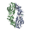
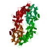
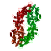
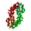
| Title | Structure of the peptidylarginine deiminase type III (PAD3) in complex with Cl-amidine | ||||||||||||
 Components Components | Protein-arginine deiminase type-3 | ||||||||||||
 Keywords Keywords | HYDROLASE / Posttransrational modification / citrullination / calcium-dependent / calcium metabolism / CYTOSOLIC PROTEIN | ||||||||||||
| Function / homology |  Function and homology information Function and homology informationprotein-arginine deiminase / protein-arginine deiminase activity / Chromatin modifying enzymes / calcium ion binding / nucleoplasm / identical protein binding / nucleus / cytoplasm / cytosol Similarity search - Function | ||||||||||||
| Biological species |  Homo sapiens (human) Homo sapiens (human) | ||||||||||||
| Method |  X-RAY DIFFRACTION / SYNCHROTRON / MOLECULAR REPLACEMENT / Resolution: 3.175 Å X-RAY DIFFRACTION / SYNCHROTRON / MOLECULAR REPLACEMENT / Resolution: 3.175 Å | ||||||||||||
 Authors Authors | Funabashi, K. / Unno, M. | ||||||||||||
| Funding support |  Japan, 3items Japan, 3items
| ||||||||||||
 Citation Citation | Journal: Arch.Biochem.Biophys. / Year: 2021 Title: Structures of human peptidylarginine deiminase type III provide insights into substrate recognition and inhibitor design. Authors: Funabashi, K. / Sawata, M. / Nagai, A. / Akimoto, M. / Mashimo, R. / Takahara, H. / Kizawa, K. / Thompson, P.R. / Ite, K. / Kitanishi, K. / Unno, M. | ||||||||||||
| History |
|
- Structure visualization
| Structure viewer | Molecule: MolmilJmol/JSmol |
|---|
- Downloads & links
-Download
| PDBx/mmCIF format | 7d56.cif.gz | 758.5 KB | Display | PDBx/mmCIF format |
|---|---|---|---|---|
| PDB format | pdb7d56.ent.gz | 633.8 KB | Display | PDB format |
| PDBx/mmJSON format | 7d56.json.gz | Tree view | PDBx/mmJSON format | |
| Others |  Other downloads Other downloads |
-Validation report
| Arichive directory | https://data.pdbj.org/pub/pdb/validation_reports/d5/7d56ftp://data.pdbj.org/pub/pdb/validation_reports/d5/7d56 | HTTPS FTP |
|---|
-Related structure data
| Related structure data |  7d4ySC 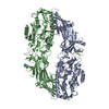 7d5rC  7d5vC 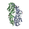 7d8nC 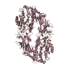 7danC S: Starting model for refinement C: citing same article ( |
|---|---|
| Similar structure data |

-Links
 PDBj
PDBj- Assembly
| Deposited unit | 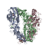
| |||||||||||||||||||||||||||||||||||||||||||||||||||||||||||||||||||||||||||||||||||||||||||||
|---|---|---|---|---|---|---|---|---|---|---|---|---|---|---|---|---|---|---|---|---|---|---|---|---|---|---|---|---|---|---|---|---|---|---|---|---|---|---|---|---|---|---|---|---|---|---|---|---|---|---|---|---|---|---|---|---|---|---|---|---|---|---|---|---|---|---|---|---|---|---|---|---|---|---|---|---|---|---|---|---|---|---|---|---|---|---|---|---|---|---|---|---|---|---|
| 1 | 
| |||||||||||||||||||||||||||||||||||||||||||||||||||||||||||||||||||||||||||||||||||||||||||||
| 2 | 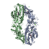
| |||||||||||||||||||||||||||||||||||||||||||||||||||||||||||||||||||||||||||||||||||||||||||||
| Unit cell |
| |||||||||||||||||||||||||||||||||||||||||||||||||||||||||||||||||||||||||||||||||||||||||||||
| Noncrystallographic symmetry (NCS) | NCS domain:
NCS domain segments: Ens-ID: 1
|