 Movie
Movie Controller

[English] 日本語
 Yorodumi
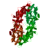
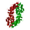
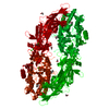
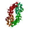
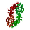
- PDB-4n2n: Crystal structure of Protein Arginine Deiminase 2 (E354A, 10 mM Ca2+) -
+ Open data

- Basic information
| Entry | Database: PDB / ID: 4n2n | ||||||
|---|---|---|---|---|---|---|---|
| Title | Crystal structure of Protein Arginine Deiminase 2 (E354A, 10 mM Ca2+) | ||||||
 Components Components | Protein-arginine deiminase type-2 | ||||||
 Keywords Keywords | HYDROLASE / deiminase | ||||||
| Function / homology |  Function and homology information Function and homology informationnegative regulation of lymphocyte chemotaxis / negative regulation of chemokine-mediated signaling pathway / histone arginine deiminase activity / histone H3R26 arginine deiminase activity / protein-arginine deiminase / protein-arginine deiminase activity / estrogen receptor signaling pathway / Chromatin modifying enzymes / substantia nigra development / transcription initiation-coupled chromatin remodeling ...negative regulation of lymphocyte chemotaxis / negative regulation of chemokine-mediated signaling pathway / histone arginine deiminase activity / histone H3R26 arginine deiminase activity / protein-arginine deiminase / protein-arginine deiminase activity / estrogen receptor signaling pathway / Chromatin modifying enzymes / substantia nigra development / transcription initiation-coupled chromatin remodeling / nuclear estrogen receptor binding / euchromatin / azurophil granule lumen / chromatin remodeling / calcium ion binding / Neutrophil degranulation / protein homodimerization activity / extracellular exosome / extracellular region / nucleus / cytosol / cytoplasm Similarity search - Function | ||||||
| Biological species |  Homo sapiens (human) Homo sapiens (human) | ||||||
| Method |  X-RAY DIFFRACTION / SYNCHROTRON / MOLECULAR REPLACEMENT / Resolution: 1.8 Å X-RAY DIFFRACTION / SYNCHROTRON / MOLECULAR REPLACEMENT / Resolution: 1.8 Å | ||||||
 Authors Authors | Slade, D.J. / Zhang, X. / Fang, P. / Dreyton, C.J. / Zhang, Y. / Gross, M.L. / Guo, M. / Coonrod, S.A. / Thompson, P.R. | ||||||
 Citation Citation | Journal: Acs Chem.Biol. / Year: 2015 Title: Protein arginine deiminase 2 binds calcium in an ordered fashion: implications for inhibitor design. Authors: Slade, D.J. / Fang, P. / Dreyton, C.J. / Zhang, Y. / Fuhrmann, J. / Rempel, D. / Bax, B.D. / Coonrod, S.A. / Lewis, H.D. / Guo, M. / Gross, M.L. / Thompson, P.R. | ||||||
| History |
|
- Structure visualization
| Structure viewer | Molecule: MolmilJmol/JSmol |
|---|
- Downloads & links
-Download
| PDBx/mmCIF format | 4n2n.cif.gz | 294.1 KB | Display | PDBx/mmCIF format |
|---|---|---|---|---|
| PDB format | pdb4n2n.ent.gz | 232.1 KB | Display | PDB format |
| PDBx/mmJSON format | 4n2n.json.gz | Tree view | PDBx/mmJSON format | |
| Others |  Other downloads Other downloads |
-Validation report
| Arichive directory | https://data.pdbj.org/pub/pdb/validation_reports/n2/4n2nftp://data.pdbj.org/pub/pdb/validation_reports/n2/4n2n | HTTPS FTP |
|---|
-Related structure data
| Related structure data |  4n20C  4n22C  4n24C  4n25SC  4n26C  4n28C  4n2aC  4n2bC  4n2cC 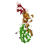 4n2dC 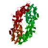 4n2eC  4n2fC 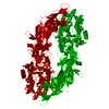 4n2gC 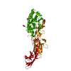 4n2hC  4n2iC  4n2kC  4n2lC 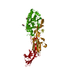 4n2mC C: citing same article ( S: Starting model for refinement |
|---|---|
| Similar structure data |


-Links
 PDBj
PDBj- Assembly
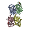
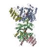
| Deposited unit | 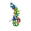
| ||||||||
|---|---|---|---|---|---|---|---|---|---|
| 1 | 
| ||||||||
| Unit cell |
| ||||||||
| Components on special symmetry positions |
|
-Components
| #1: Protein | Mass: 78614.969 Da / Num. of mol.: 1 / Mutation: E354A Source method: isolated from a genetically manipulated source Source: (gene. exp.) Homo sapiens (human) / Gene: PADI2, KIAA0994, PDI2 / Production host:  | ||||||
|---|---|---|---|---|---|---|---|
| #2: Chemical |   Mass: 118.174 Da / Num. of mol.: 3 / Source method: obtained synthetically / Formula: C6H14O2 / Comment: precipitant*YM Mass: 118.174 Da / Num. of mol.: 3 / Source method: obtained synthetically / Formula: C6H14O2 / Comment: precipitant*YM#3: Chemical | ChemComp-CA /   Mass: 40.078 Da / Num. of mol.: 4 / Source method: obtained synthetically / Formula: Ca Mass: 40.078 Da / Num. of mol.: 4 / Source method: obtained synthetically / Formula: Ca#4: Chemical |   Mass: 59.044 Da / Num. of mol.: 2 / Source method: obtained synthetically / Formula: C2H3O2 Mass: 59.044 Da / Num. of mol.: 2 / Source method: obtained synthetically / Formula: C2H3O2#5: Water | ChemComp-HOH / |  Mass: 18.015 Da / Num. of mol.: 653 / Source method: isolated from a natural source / Formula: H2O Mass: 18.015 Da / Num. of mol.: 653 / Source method: isolated from a natural source / Formula: H2O |
-Experimental details
-Experiment
| Experiment | Method: X-RAY DIFFRACTION / Number of used crystals: 1 |
|---|
- Sample preparation
| Crystal | Density Matthews: 2.45 Å3/Da / Density % sol: 49.88 % |
|---|---|
| Crystal grow | Method: vapor diffusion, sitting drop / pH: 5.6 Details: 10-20% MPD, 50 mM MES, pH 5.6, 0.12 M sodium acetate, VAPOR DIFFUSION, SITTING DROP |
-Data collection
| Diffraction | Mean temperature: 100 K |
|---|---|
| Diffraction source | Source: SYNCHROTRON / Site: APS  / Beamline: 21-ID-G / Wavelength: 0.9787 Å / Beamline: 21-ID-G / Wavelength: 0.9787 Å |
| Detector | Type: MARMOSAIC 300 mm CCD / Detector: CCD / Date: Jan 1, 2012 / Details: beryllium lenses |
| Radiation | Monochromator: diamond(111) / Protocol: SINGLE WAVELENGTH / Monochromatic (M) / Laue (L): M / Scattering type: x-ray |
| Radiation wavelength | Wavelength: 0.9787 Å / Relative weight: 1 |
| Reflection | Resolution: 1.8→38.063 Å / Num. obs: 70612 / % possible obs: 99.5 % / Redundancy: 4.2 % / Biso Wilson estimate: 22.06 Å2 / Rmerge(I) obs: 0.094 / Rsym value: 0.081 |
| Reflection shell | Highest resolution: 1.8 Å / Rmerge(I) obs: 0.536 / Mean I/σ(I) obs: 2.3 / % possible all: 99 |
- Processing
| Software |
| |||||||||||||||||||||||||||||||||||||||||||||||||||||||||||||||||||||||||||||
|---|---|---|---|---|---|---|---|---|---|---|---|---|---|---|---|---|---|---|---|---|---|---|---|---|---|---|---|---|---|---|---|---|---|---|---|---|---|---|---|---|---|---|---|---|---|---|---|---|---|---|---|---|---|---|---|---|---|---|---|---|---|---|---|---|---|---|---|---|---|---|---|---|---|---|---|---|---|---|
| Refinement | Method to determine structure: MOLECULAR REPLACEMENT Starting model: PDB ENTRY 4N25 Resolution: 1.8→38.063 Å / Occupancy max: 1 / Occupancy min: 0.24 / FOM work R set: 0.8658 / SU ML: 0.5 / σ(F): 1.34 / Phase error: 20.52 / Stereochemistry target values: ML
| |||||||||||||||||||||||||||||||||||||||||||||||||||||||||||||||||||||||||||||
| Solvent computation | Shrinkage radii: 0.9 Å / VDW probe radii: 1.11 Å / Solvent model: FLAT BULK SOLVENT MODEL / Bsol: 45.662 Å2 / ksol: 0.346 e/Å3 | |||||||||||||||||||||||||||||||||||||||||||||||||||||||||||||||||||||||||||||
| Displacement parameters | Biso max: 101.99 Å2 / Biso mean: 29.3074 Å2 / Biso min: 9.81 Å2
| |||||||||||||||||||||||||||||||||||||||||||||||||||||||||||||||||||||||||||||
| Refinement step | Cycle: LAST / Resolution: 1.8→38.063 Å
| |||||||||||||||||||||||||||||||||||||||||||||||||||||||||||||||||||||||||||||
| Refine LS restraints |
| |||||||||||||||||||||||||||||||||||||||||||||||||||||||||||||||||||||||||||||
| LS refinement shell | Refine-ID: X-RAY DIFFRACTION / Total num. of bins used: 10
|
