 Movie
Movie Controller

[English] 日本語
 Yorodumi
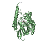
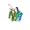
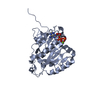
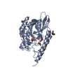
- PDB-7d18: Crystal structure of Acidobacteriales bacterium glutaminyl cyclase -
+ Open data

- Basic information
| Entry | Database: PDB / ID: 7d18 | ||||||
|---|---|---|---|---|---|---|---|
| Title | Crystal structure of Acidobacteriales bacterium glutaminyl cyclase | ||||||
 Components Components | Peptidase M28 | ||||||
 Keywords Keywords | TRANSFERASE / Glutaminyl cyclase / METAL BINDING PROTEIN | ||||||
| Function / homology | Glutaminyl-peptide cyclotransferase-like / glutaminyl-peptide cyclotransferase activity / Peptidase M28 / Peptidase family M28 / zinc ion binding / Peptidase M28 Function and homology information Function and homology information | ||||||
| Biological species |  Acidobacteriales bacterium 59-55 (bacteria) Acidobacteriales bacterium 59-55 (bacteria) | ||||||
| Method |  X-RAY DIFFRACTION / SYNCHROTRON / SAD / Resolution: 1.332 Å X-RAY DIFFRACTION / SYNCHROTRON / SAD / Resolution: 1.332 Å | ||||||
 Authors Authors | Huang, K.-F. / Huang, J.-S. / Wu, M.-L. / Hsieh, W.-L. / Wang, A.H.-J. | ||||||
| Funding support |  Taiwan, 1items Taiwan, 1items
| ||||||
 Citation Citation | Journal: J.Mol.Biol. / Year: 2021 Title: A Unique Carboxylic-Acid Hydrogen-Bond Network (CAHBN) Confers Glutaminyl Cyclase Activity on M28 Family Enzymes. Authors: Huang, K.F. / Huang, J.S. / Wu, M.L. / Hsieh, W.L. / Hsu, K.C. / Hsu, H.L. / Ko, T.P. / Wang, A.H. | ||||||
| History |
|
- Structure visualization
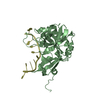
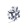
| Structure viewer | Molecule: MolmilJmol/JSmol |
|---|
- Downloads & links
-Download
| PDBx/mmCIF format | 7d18.cif.gz | 146.9 KB | Display | PDBx/mmCIF format |
|---|---|---|---|---|
| PDB format | pdb7d18.ent.gz | 113.4 KB | Display | PDB format |
| PDBx/mmJSON format | 7d18.json.gz | Tree view | PDBx/mmJSON format | |
| Others |  Other downloads Other downloads |
-Validation report
| Arichive directory | https://data.pdbj.org/pub/pdb/validation_reports/d1/7d18ftp://data.pdbj.org/pub/pdb/validation_reports/d1/7d18 | HTTPS FTP |
|---|
-Related structure data
| Related structure data |  7d17C 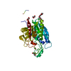 7d1bC  7d1dC 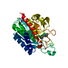 7d1eC  7d1hC  7d1nC  7d1pC  7d1yC  7d21C  7d23C  7d2bC  7d2dC  7d2iC  7d2jC C: citing same article ( |
|---|---|
| Similar structure data |


-Links
 PDBj
PDBj
- Assembly
| Deposited unit | 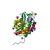
| ||||||||
|---|---|---|---|---|---|---|---|---|---|
| 1 |
| ||||||||
| Unit cell |
|
-Components
-Protein , 1 types, 1 molecules A
| #1: Protein | Mass: 32564.648 Da / Num. of mol.: 1 Source method: isolated from a genetically manipulated source Source: (gene. exp.) Acidobacteriales bacterium 59-55 (bacteria)Gene: BGO25_06485 / Production host: |
|---|
-Non-polymers , 6 types, 346 molecules 


| #2: Chemical | ChemComp-ZN /  Mass: 65.409 Da / Num. of mol.: 1 / Source method: obtained synthetically / Formula: Zn Mass: 65.409 Da / Num. of mol.: 1 / Source method: obtained synthetically / Formula: Zn | ||||||
|---|---|---|---|---|---|---|---|
| #3: Chemical | ChemComp-NA /  Mass: 22.990 Da / Num. of mol.: 1 / Source method: obtained synthetically / Formula: Na Mass: 22.990 Da / Num. of mol.: 1 / Source method: obtained synthetically / Formula: Na | ||||||
| #4: Chemical | ChemComp-SO4 /  Mass: 96.063 Da / Num. of mol.: 7 / Source method: obtained synthetically / Formula: SO4 Mass: 96.063 Da / Num. of mol.: 7 / Source method: obtained synthetically / Formula: SO4#5: Chemical | ChemComp-GOL /  Mass: 92.094 Da / Num. of mol.: 5 / Source method: obtained synthetically / Formula: C3H8O3 Mass: 92.094 Da / Num. of mol.: 5 / Source method: obtained synthetically / Formula: C3H8O3#6: Chemical | ChemComp-TRS / |  Mass: 122.143 Da / Num. of mol.: 1 / Source method: obtained synthetically / Formula: C4H12NO3 / Comment: pH buffer*YM Mass: 122.143 Da / Num. of mol.: 1 / Source method: obtained synthetically / Formula: C4H12NO3 / Comment: pH buffer*YM#7: Water | ChemComp-HOH / | Mass: 18.015 Da / Num. of mol.: 331 / Source method: isolated from a natural source / Formula: H2O |
-Details
| Has ligand of interest | N |
|---|
-Experimental details
-Experiment
| Experiment | Method: X-RAY DIFFRACTION / Number of used crystals: 1 |
|---|
- Sample preparation
| Crystal | Density Matthews: 1.87 Å3/Da / Density % sol: 31.15 % |
|---|---|
| Crystal grow | Temperature: 293.15 K / Method: vapor diffusion, hanging drop / pH: 6.5 Details: 0.2 M (NH4)2SO4, 0.1 M sodium cacodylate, pH 6.5, 30% (w/v) PEG 8000 |
-Data collection
| Diffraction | Mean temperature: 100 K / Serial crystal experiment: N |
|---|---|
| Diffraction source | Source: SYNCHROTRON / Site: NSRRC / Beamline: BL15A1 / Wavelength: 1 Å |
| Detector | Type: RAYONIX MX300HE / Detector: CCD / Date: Aug 1, 2019 |
| Radiation | Protocol: SINGLE WAVELENGTH / Monochromatic (M) / Laue (L): M / Scattering type: x-ray |
| Radiation wavelength | Wavelength: 1 Å / Relative weight: 1 |
| Reflection | Resolution: 1.33→30 Å / Num. obs: 53659 / % possible obs: 98.5 % / Redundancy: 10 % / Biso Wilson estimate: 13.73 Å2 / Rmerge(I) obs: 0.107 / Net I/σ(I): 28 |
| Reflection shell | Resolution: 1.33→1.38 Å / Redundancy: 7.3 % / Rmerge(I) obs: 0.775 / Mean I/σ(I) obs: 3.9 / Num. unique obs: 5037 / % possible all: 92.6 |
-Phasing
| Phasing | Method: SAD |
|---|
- Processing
| Software |
| ||||||||||||||||||||||||||||||||||||||||||||||||||||||||||||||||||||||||||||||||||||||||||
|---|---|---|---|---|---|---|---|---|---|---|---|---|---|---|---|---|---|---|---|---|---|---|---|---|---|---|---|---|---|---|---|---|---|---|---|---|---|---|---|---|---|---|---|---|---|---|---|---|---|---|---|---|---|---|---|---|---|---|---|---|---|---|---|---|---|---|---|---|---|---|---|---|---|---|---|---|---|---|---|---|---|---|---|---|---|---|---|---|---|---|---|
| Refinement | Method to determine structure: SAD / Resolution: 1.332→25.909 Å / SU ML: 0.1 / Cross valid method: THROUGHOUT / σ(F): 1.35 / Phase error: 11.86 / Stereochemistry target values: ML
| ||||||||||||||||||||||||||||||||||||||||||||||||||||||||||||||||||||||||||||||||||||||||||
| Solvent computation | Shrinkage radii: 0.9 Å / VDW probe radii: 1.11 Å / Solvent model: FLAT BULK SOLVENT MODEL | ||||||||||||||||||||||||||||||||||||||||||||||||||||||||||||||||||||||||||||||||||||||||||
| Displacement parameters | Biso max: 63.04 Å2 / Biso mean: 18.5238 Å2 / Biso min: 9.5 Å2 | ||||||||||||||||||||||||||||||||||||||||||||||||||||||||||||||||||||||||||||||||||||||||||
| Refinement step | Cycle: final / Resolution: 1.332→25.909 Å
| ||||||||||||||||||||||||||||||||||||||||||||||||||||||||||||||||||||||||||||||||||||||||||
| LS refinement shell | Refine-ID: X-RAY DIFFRACTION / Rfactor Rfree error: 0
|
