 Movie
Movie Controller

[English] 日本語
 Yorodumi
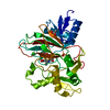
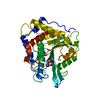
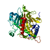
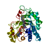
- PDB-7d21: Crystal structure of Ixodes scapularis glutaminyl cyclase with tw... -
+ Open data

- Basic information
| Entry | Database: PDB / ID: 7d21 | ||||||
|---|---|---|---|---|---|---|---|
| Title | Crystal structure of Ixodes scapularis glutaminyl cyclase with two Zn ions bound to the active site | ||||||
 Components Components | Glutaminyl-peptide cyclotransferase | ||||||
 Keywords Keywords | TRANSFERASE / Glutaminyl cyclase / METAL BINDING PROTEIN | ||||||
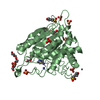
| Function / homology |  Function and homology information Function and homology informationpeptidyl-pyroglutamic acid biosynthetic process, using glutaminyl-peptide cyclotransferase / glutaminyl-peptide cyclotransferase / glutaminyl-peptide cyclotransferase activity / extracellular region / zinc ion binding Similarity search - Function | ||||||
| Biological species |  Ixodes scapularis (black-legged tick) Ixodes scapularis (black-legged tick) | ||||||
| Method |  X-RAY DIFFRACTION / SYNCHROTRON / MOLECULAR REPLACEMENT / molecular replacement / Resolution: 1.97 Å X-RAY DIFFRACTION / SYNCHROTRON / MOLECULAR REPLACEMENT / molecular replacement / Resolution: 1.97 Å | ||||||
 Authors Authors | Huang, K.-F. / Huang, J.-S. / Wu, M.-L. / Hsieh, W.-L. / Wang, A.H.-J. | ||||||
| Funding support |  Taiwan, 1items Taiwan, 1items
| ||||||
 Citation Citation | Journal: J.Mol.Biol. / Year: 2021 Title: A Unique Carboxylic-Acid Hydrogen-Bond Network (CAHBN) Confers Glutaminyl Cyclase Activity on M28 Family Enzymes. Authors: Huang, K.F. / Huang, J.S. / Wu, M.L. / Hsieh, W.L. / Hsu, K.C. / Hsu, H.L. / Ko, T.P. / Wang, A.H. | ||||||
| History |
|
- Structure visualization
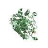
| Structure viewer | Molecule: MolmilJmol/JSmol |
|---|
- Downloads & links
-Download
| PDBx/mmCIF format | 7d21.cif.gz | 87 KB | Display | PDBx/mmCIF format |
|---|---|---|---|---|
| PDB format | pdb7d21.ent.gz | 62.8 KB | Display | PDB format |
| PDBx/mmJSON format | 7d21.json.gz | Tree view | PDBx/mmJSON format | |
| Others |  Other downloads Other downloads |
-Validation report
| Arichive directory | https://data.pdbj.org/pub/pdb/validation_reports/d2/7d21ftp://data.pdbj.org/pub/pdb/validation_reports/d2/7d21 | HTTPS FTP |
|---|
-Related structure data
| Related structure data |  7d17C  7d18C  7d1bC  7d1dC  7d1eC  7d1hC  7d1nC  7d1pC  7d1yC  7d23C  7d2bC  7d2dC  7d2iC  7d2jC  4mhnS S: Starting model for refinement C: citing same article ( |
|---|---|
| Similar structure data |


-Links
 PDBj
PDBj
- Assembly
| Deposited unit | 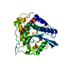
| ||||||||
|---|---|---|---|---|---|---|---|---|---|
| 1 |
| ||||||||
| Unit cell |
|
-Components
| #1: Protein | Mass: 38248.105 Da / Num. of mol.: 1 Source method: isolated from a genetically manipulated source Source: (gene. exp.) Ixodes scapularis (black-legged tick) / Gene: 8042451, IscW_ISCW023264 / Production host:  References: UniProt: B7QK46, glutaminyl-peptide cyclotransferase | ||||||||
|---|---|---|---|---|---|---|---|---|---|
| #2: Chemical | ChemComp-ZN /   Mass: 65.409 Da / Num. of mol.: 5 / Source method: obtained synthetically / Formula: Zn Mass: 65.409 Da / Num. of mol.: 5 / Source method: obtained synthetically / Formula: Zn#3: Chemical | ChemComp-EPE / |   Mass: 238.305 Da / Num. of mol.: 1 / Source method: obtained synthetically / Formula: C8H18N2O4S / Comment: pH buffer*YM Mass: 238.305 Da / Num. of mol.: 1 / Source method: obtained synthetically / Formula: C8H18N2O4S / Comment: pH buffer*YM#4: Water | ChemComp-HOH / |  Mass: 18.015 Da / Num. of mol.: 146 / Source method: isolated from a natural source / Formula: H2O Mass: 18.015 Da / Num. of mol.: 146 / Source method: isolated from a natural source / Formula: H2OHas ligand of interest | N | Has protein modification | Y | |
-Experimental details
-Experiment
| Experiment | Method: X-RAY DIFFRACTION / Number of used crystals: 1 |
|---|
- Sample preparation
| Crystal | Density Matthews: 2.11 Å3/Da / Density % sol: 41.61 % |
|---|---|
| Crystal grow | Temperature: 293.15 K / Method: vapor diffusion, sitting drop / pH: 7.5 Details: 10%(w/v) PEG8000, 8%(v/v) ethylene glycol, 0.1 M HEPES pH 7.5 |
-Data collection
| Diffraction | Mean temperature: 100 K / Serial crystal experiment: N |
|---|---|
| Diffraction source | Source: SYNCHROTRON / Site: NSRRC / Beamline: BL15A1 / Wavelength: 1 Å |
| Detector | Type: RAYONIX MX300HE / Detector: CCD / Date: Oct 10, 2017 |
| Radiation | Protocol: SINGLE WAVELENGTH / Monochromatic (M) / Laue (L): M / Scattering type: x-ray |
| Radiation wavelength | Wavelength: 1 Å / Relative weight: 1 |
| Reflection | Resolution: 1.97→30 Å / Num. obs: 23206 / % possible obs: 99.9 % / Redundancy: 5.6 % / Rmerge(I) obs: 0.09 / Net I/σ(I): 31.1 |
| Reflection shell | Resolution: 1.97→2.04 Å / Redundancy: 5.6 % / Rmerge(I) obs: 0.855 / Mean I/σ(I) obs: 2.6 / Num. unique obs: 2278 / % possible all: 100 |
-Phasing
| Phasing | Method: molecular replacement |
|---|
- Processing
| Software |
| ||||||||||||||||||||||||||||||||||||||||||||||||||||||||||||
|---|---|---|---|---|---|---|---|---|---|---|---|---|---|---|---|---|---|---|---|---|---|---|---|---|---|---|---|---|---|---|---|---|---|---|---|---|---|---|---|---|---|---|---|---|---|---|---|---|---|---|---|---|---|---|---|---|---|---|---|---|---|
| Refinement | Method to determine structure: MOLECULAR REPLACEMENT Starting model: 4MHN Resolution: 1.97→27.84 Å / Cor.coef. Fo:Fc: 0.959 / Cor.coef. Fo:Fc free: 0.949 / SU B: 3.902 / SU ML: 0.111 / Cross valid method: THROUGHOUT / σ(F): 0 / ESU R: 0.197 / ESU R Free: 0.161 / Stereochemistry target values: MAXIMUM LIKELIHOOD Details: HYDROGENS HAVE BEEN ADDED IN THE RIDING POSITIONS U VALUES : REFINED INDIVIDUALLY
| ||||||||||||||||||||||||||||||||||||||||||||||||||||||||||||
| Solvent computation | Ion probe radii: 0.8 Å / Shrinkage radii: 0.8 Å / VDW probe radii: 1.2 Å / Solvent model: MASK | ||||||||||||||||||||||||||||||||||||||||||||||||||||||||||||
| Displacement parameters | Biso max: 114.13 Å2 / Biso mean: 41.856 Å2 / Biso min: 21.36 Å2
| ||||||||||||||||||||||||||||||||||||||||||||||||||||||||||||
| Refinement step | Cycle: final / Resolution: 1.97→27.84 Å
| ||||||||||||||||||||||||||||||||||||||||||||||||||||||||||||
| Refine LS restraints |
| ||||||||||||||||||||||||||||||||||||||||||||||||||||||||||||
| LS refinement shell | Resolution: 1.97→2.019 Å / Rfactor Rfree error: 0
|
