 Movie
Movie Controller

[English] 日本語
 Yorodumi
- PDB-7czb: The cryo-EM structure of the ERAD retrotranslocation channel form... -
+ Open data

- Basic information
| Entry | Database: PDB / ID: 7czb | |||||||||||||||
|---|---|---|---|---|---|---|---|---|---|---|---|---|---|---|---|---|
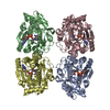
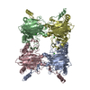
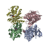
| Title | The cryo-EM structure of the ERAD retrotranslocation channel formed by human Derlin-1 | |||||||||||||||
 Components Components | Derlin-1 | |||||||||||||||
 Keywords Keywords | MEMBRANE PROTEIN / Endoplasmic reticulum associated degradation / Retrotranslocation / cryo-EM / Transmembrane channels | |||||||||||||||
| Function / homology |  Function and homology information Function and homology informationDerlin-1-VIMP complex / signal recognition particle binding / endoplasmic reticulum quality control compartment / Derlin-1 retrotranslocation complex / cellular response to misfolded protein / retrograde protein transport, ER to cytosol / ubiquitin-specific protease binding / MHC class I protein binding / response to unfolded protein / endoplasmic reticulum unfolded protein response ...Derlin-1-VIMP complex / signal recognition particle binding / endoplasmic reticulum quality control compartment / Derlin-1 retrotranslocation complex / cellular response to misfolded protein / retrograde protein transport, ER to cytosol / ubiquitin-specific protease binding / MHC class I protein binding / response to unfolded protein / endoplasmic reticulum unfolded protein response / ERAD pathway / positive regulation of protein ubiquitination / N-glycan trimming in the ER and Calnexin/Calreticulin cycle / establishment of protein localization / Defective CFTR causes cystic fibrosis / protein destabilization / ABC-family protein mediated transport / late endosome / E3 ubiquitin ligases ubiquitinate target proteins / ATPase binding / signaling receptor activity / protease binding / proteasome-mediated ubiquitin-dependent protein catabolic process / early endosome / ubiquitin protein ligase binding / endoplasmic reticulum membrane / protein-containing complex binding / endoplasmic reticulum / membrane / identical protein binding Similarity search - Function | |||||||||||||||
| Biological species |  Homo sapiens (human) Homo sapiens (human) | |||||||||||||||
| Method | ELECTRON MICROSCOPY / single particle reconstruction / cryo EM / Resolution: 3.8 Å | |||||||||||||||
 Authors Authors | Rao, B. / Li, S. / Yao, D. / Wang, Q. / Xia, Y. / Jia, Y. / Shen, Y. / Cao, Y. | |||||||||||||||
| Funding support |  China, 4items China, 4items
| |||||||||||||||
 Citation Citation | Journal: Sci Adv / Year: 2021 Title: The cryo-EM structure of an ERAD protein channel formed by tetrameric human Derlin-1. Authors: Bing Rao / Shaobai Li / Deqiang Yao / Qian Wang / Ying Xia / Yi Jia / Yafeng Shen / Yu Cao / Abstract: Endoplasmic reticulum-associated degradation (ERAD) is a process directing misfolded proteins from the ER lumen and membrane to the degradation machinery in the cytosol. A key step in ERAD is the ...Endoplasmic reticulum-associated degradation (ERAD) is a process directing misfolded proteins from the ER lumen and membrane to the degradation machinery in the cytosol. A key step in ERAD is the translocation of ER proteins to the cytosol. Derlins are essential for protein translocation in ERAD, but the mechanism remains unclear. Here, we solved the structure of human Derlin-1 by cryo-electron microscopy. The structure shows that Derlin-1 forms a homotetramer that encircles a large tunnel traversing the ER membrane. The tunnel has a diameter of about 12 to 15 angstroms, large enough to allow an α helix to pass through. The structure also shows a lateral gate within the membrane, providing access of transmembrane proteins to the tunnel, and thus, human Derlin-1 forms a protein channel for translocation of misfolded proteins. Our structure is different from the monomeric yeast Derlin structure previously reported, which forms a semichannel with another protein. | |||||||||||||||
| History |
|
- Structure visualization
| Movie |
Movie viewer |
|---|---|
| Structure viewer | Molecule: MolmilJmol/JSmol |
- Downloads & links
-Download
| PDBx/mmCIF format | 7czb.cif.gz | 146.7 KB | Display | PDBx/mmCIF format |
|---|---|---|---|---|
| PDB format | pdb7czb.ent.gz | 116.3 KB | Display | PDB format |
| PDBx/mmJSON format | 7czb.json.gz | Tree view | PDBx/mmJSON format | |
| Others |  Other downloads Other downloads |
-Validation report
| Arichive directory | https://data.pdbj.org/pub/pdb/validation_reports/cz/7czbftp://data.pdbj.org/pub/pdb/validation_reports/cz/7czb | HTTPS FTP |
|---|
-Related structure data
| Related structure data |  30508MC M: map data used to model this data C: citing same article ( |
|---|---|
| Similar structure data |


-Links
 PDBj
PDBj


- Assembly
| Deposited unit | 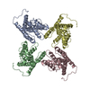
|
|---|---|
| 1 |
|
-Components
| #1: Protein | Mass: 28821.664 Da / Num. of mol.: 4 Source method: isolated from a genetically manipulated source Source: (gene. exp.) Homo sapiens (human) / Gene: DERL1, DER1, UNQ243/PRO276 / Plasmid: pcDNA3.4 / Cell line (production host): Expi293F / Production host: Homo sapiens (human) / References: UniProt: Q9BUN8 |
|---|
-Experimental details
-Experiment
| Experiment | Method: ELECTRON MICROSCOPY |
|---|---|
| EM experiment | Aggregation state: PARTICLE / 3D reconstruction method: single particle reconstruction |
- Sample preparation
| Component | Name: Derlin-1 / Type: CELL / Entity ID: all / Source: RECOMBINANT |
|---|---|
| Source (natural) | Organism: Homo sapiens (human) |
| Source (recombinant) | Organism: Homo sapiens (human) / Cell: Expi293F / Plasmid: pcDNA3.4 |
| Buffer solution | pH: 8 |
| Specimen | Conc.: 1.5 mg/ml / Embedding applied: NO / Shadowing applied: NO / Staining applied: NO / Vitrification applied: YES |
| Specimen support | Grid material: GOLD / Grid mesh size: 300 divisions/in. / Grid type: Quantifoil R1.2/1.3 |
| Vitrification | Instrument: FEI VITROBOT MARK IV / Cryogen name: ETHANE / Humidity: 100 % / Chamber temperature: 283.15 K |
- Electron microscopy imaging
| Experimental equipment |  Model: Titan Krios / Image courtesy: FEI Company |
|---|---|
| Microscopy | Model: FEI TITAN KRIOS |
| Electron gun | Electron source:  FIELD EMISSION GUN / Accelerating voltage: 300 kV / Illumination mode: FLOOD BEAM FIELD EMISSION GUN / Accelerating voltage: 300 kV / Illumination mode: FLOOD BEAM |
| Electron lens | Mode: BRIGHT FIELD / Alignment procedure: COMA FREE |
| Specimen holder | Cryogen: NITROGEN / Specimen holder model: FEI TITAN KRIOS AUTOGRID HOLDER |
| Image recording | Average exposure time: 2.7 sec. / Electron dose: 50 e/Å2 / Film or detector model: GATAN K3 BIOQUANTUM (6k x 4k) |
- Processing
| CTF correction | Type: PHASE FLIPPING AND AMPLITUDE CORRECTION |
|---|---|
| Symmetry | Point symmetry: C2 (2 fold cyclic) |
| 3D reconstruction | Resolution: 3.8 Å / Resolution method: FSC 0.143 CUT-OFF / Num. of particles: 71761 / Algorithm: FOURIER SPACE / Symmetry type: POINT |
