 Movie
Movie Controller

[English] 日本語
 Yorodumi
- PDB-1rkx: Crystal Structure at 1.8 Angstrom of CDP-D-glucose 4,6-dehydratas... -
+ Open data

- Basic information
| Entry | Database: PDB / ID: 1rkx | ||||||
|---|---|---|---|---|---|---|---|
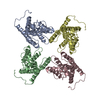
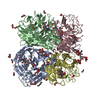
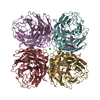
| Title | Crystal Structure at 1.8 Angstrom of CDP-D-glucose 4,6-dehydratase from Yersinia pseudotuberculosis | ||||||
 Components Components | CDP-glucose-4,6-dehydratase | ||||||
 Keywords Keywords | LYASE / Dehydratase / SDR / CDP Glucose dehydratase | ||||||
| Function / homology |  Function and homology information Function and homology informationCDP-glucose 4,6-dehydratase / CDP-glucose 4,6-dehydratase activity / nucleotide binding Similarity search - Function | ||||||
| Biological species |  Yersinia pseudotuberculosis (bacteria) Yersinia pseudotuberculosis (bacteria) | ||||||
| Method |  X-RAY DIFFRACTION / SYNCHROTRON / MAD, MIR / Resolution: 1.8 Å X-RAY DIFFRACTION / SYNCHROTRON / MAD, MIR / Resolution: 1.8 Å | ||||||
 Authors Authors | Vogan, E.M. / Bellamacina, C. / He, X. / Liu, H.W. / Ringe, D. / Petsko, G.A. | ||||||
 Citation Citation | Journal: Biochemistry / Year: 2004 Title: Crystal Structure at 1.8 A Resolution of CDP-d-Glucose 4,6-Dehydratase from Yersinia pseudotuberculosis Authors: Vogan, E.M. / Bellamacina, C. / He, X. / Liu, H.W. / Ringe, D. / Petsko, G.A. #1: Journal: Acta Crystallogr.,Sect.D / Year: 2002Title: Purification, crystallization and molecular symmetry of CDP-D-glucose 4,6-dehydratase from Yersinia pseudotuberculosis. Authors: Vogan, E.M. / Bellamacina, C.R. / He, X. / Liu, H.-W. / Ringe, D. / Petsko, G.A. #2: Journal: To be PublishedTitle: The X-ray crystal structure of CDP-D-glucose 4,6-dehydratase from Yersinia pseduotuberculosis. | ||||||
| History |
|
- Structure visualization
| Structure viewer | Molecule: MolmilJmol/JSmol |
|---|
- Downloads & links
-Download
| PDBx/mmCIF format | 1rkx.cif.gz | 301.9 KB | Display | PDBx/mmCIF format |
|---|---|---|---|---|
| PDB format | pdb1rkx.ent.gz | 244.8 KB | Display | PDB format |
| PDBx/mmJSON format | 1rkx.json.gz | Tree view | PDBx/mmJSON format | |
| Others |  Other downloads Other downloads |
-Validation report
| Arichive directory | https://data.pdbj.org/pub/pdb/validation_reports/rk/1rkxftp://data.pdbj.org/pub/pdb/validation_reports/rk/1rkx | HTTPS FTP |
|---|
-Related structure data
| Similar structure data |
|---|


-Links
 PDBj
PDBj- Assembly
| Deposited unit | 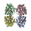
| ||||||||
|---|---|---|---|---|---|---|---|---|---|
| 1 |
| ||||||||
| Unit cell |
| ||||||||
| Details | The biological assembly is a 222 tetramer. All molecules all included explicitly. |
-Components
| #1: Protein | Mass: 40315.230 Da / Num. of mol.: 4 Source method: isolated from a genetically manipulated source Source: (gene. exp.) Yersinia pseudotuberculosis (bacteria) / Gene: ascB / Plasmid: pJT8 / Production host: #2: Chemical | ChemComp-NAD /   Mass: 663.425 Da / Num. of mol.: 4 / Source method: obtained synthetically / Formula: C21H27N7O14P2 / Comment: NAD*YM Mass: 663.425 Da / Num. of mol.: 4 / Source method: obtained synthetically / Formula: C21H27N7O14P2 / Comment: NAD*YM#3: Water | ChemComp-HOH / |  Mass: 18.015 Da / Num. of mol.: 862 / Source method: isolated from a natural source / Formula: H2O Mass: 18.015 Da / Num. of mol.: 862 / Source method: isolated from a natural source / Formula: H2O |
|---|
-Experimental details
-Experiment
| Experiment | Method: X-RAY DIFFRACTION / Number of used crystals: 1 |
|---|
- Sample preparation
| Crystal | Density Matthews: 2.3 Å3/Da / Density % sol: 46 % |
|---|---|
| Crystal grow | Temperature: 277 K / Method: vapor diffusion, hanging drop / pH: 7.5 Details: ammonium sulfate, PEG 4000, potassium HEPES, pH 7.5, VAPOR DIFFUSION, HANGING DROP, temperature 277K |
-Data collection
| Diffraction | Mean temperature: 113 K |
|---|---|
| Diffraction source | Source: SYNCHROTRON / Site: NSLS  / Beamline: X12B / Wavelength: 1.0704 Å / Beamline: X12B / Wavelength: 1.0704 Å |
| Detector | Type: ADSC QUANTUM 4 / Detector: CCD / Date: Mar 5, 1999 |
| Radiation | Monochromator: Si 111 Crystal / Protocol: SINGLE WAVELENGTH / Monochromatic (M) / Laue (L): M / Scattering type: x-ray |
| Radiation wavelength | Wavelength: 1.0704 Å / Relative weight: 1 |
| Reflection | Resolution: 1.8→50 Å / Num. all: 136502 / Num. obs: 133360 / % possible obs: 97.7 % / Observed criterion σ(F): 0 / Observed criterion σ(I): 2 / Redundancy: 5.8 % / Biso Wilson estimate: 19.8 Å2 / Rsym value: 0.066 / Net I/σ(I): 18.4 |
| Reflection shell | Resolution: 1.8→1.86 Å / Redundancy: 3.2 % / Mean I/σ(I) obs: 2.6 / Num. unique all: 13502 / Rsym value: 0.35 / % possible all: 93.4 |
- Processing
| Software |
| |||||||||||||||||||||||||||||||||||||||||||||||||||||||||||||||||||||||||||||||||||||||||||||||||||
|---|---|---|---|---|---|---|---|---|---|---|---|---|---|---|---|---|---|---|---|---|---|---|---|---|---|---|---|---|---|---|---|---|---|---|---|---|---|---|---|---|---|---|---|---|---|---|---|---|---|---|---|---|---|---|---|---|---|---|---|---|---|---|---|---|---|---|---|---|---|---|---|---|---|---|---|---|---|---|---|---|---|---|---|---|---|---|---|---|---|---|---|---|---|---|---|---|---|---|---|---|
| Refinement | Method to determine structure: MAD, MIR Starting model: NONE Resolution: 1.8→36.25 Å / Rfactor Rfree error: 0.004 / Data cutoff high absF: 4589 / Data cutoff low absF: 0 / Isotropic thermal model: RESTRAINED INDIVIDUAL / Cross valid method: THROUGHOUT / σ(F): 0 / Stereochemistry target values: Engh & Huber
| |||||||||||||||||||||||||||||||||||||||||||||||||||||||||||||||||||||||||||||||||||||||||||||||||||
| Solvent computation | Solvent model: FLAT MODEL / Bsol: 46.66 Å2 / ksol: 0.367491 e/Å3 | |||||||||||||||||||||||||||||||||||||||||||||||||||||||||||||||||||||||||||||||||||||||||||||||||||
| Displacement parameters | Biso mean: 27 Å2
| |||||||||||||||||||||||||||||||||||||||||||||||||||||||||||||||||||||||||||||||||||||||||||||||||||
| Refine analyze |
| |||||||||||||||||||||||||||||||||||||||||||||||||||||||||||||||||||||||||||||||||||||||||||||||||||
| Refinement step | Cycle: LAST / Resolution: 1.8→36.25 Å
| |||||||||||||||||||||||||||||||||||||||||||||||||||||||||||||||||||||||||||||||||||||||||||||||||||
| Refine LS restraints |
| |||||||||||||||||||||||||||||||||||||||||||||||||||||||||||||||||||||||||||||||||||||||||||||||||||
| Refine LS restraints NCS | NCS model details: RESTR | |||||||||||||||||||||||||||||||||||||||||||||||||||||||||||||||||||||||||||||||||||||||||||||||||||
| LS refinement shell | Refine-ID: X-RAY DIFFRACTION / Total num. of bins used: 10
| |||||||||||||||||||||||||||||||||||||||||||||||||||||||||||||||||||||||||||||||||||||||||||||||||||
| Xplor file |
|
