Derlin-1-VIMP complex / signal recognition particle binding / endoplasmic reticulum quality control compartment / Derlin-1 retrotranslocation complex / cellular response to misfolded protein / retrograde protein transport, ER to cytosol / ubiquitin-specific protease binding / MHC class I protein binding / response to unfolded protein / endoplasmic reticulum unfolded protein response ...Derlin-1-VIMP complex / signal recognition particle binding / endoplasmic reticulum quality control compartment / Derlin-1 retrotranslocation complex / cellular response to misfolded protein / retrograde protein transport, ER to cytosol / ubiquitin-specific protease binding / MHC class I protein binding / response to unfolded protein / endoplasmic reticulum unfolded protein response / ERAD pathway / positive regulation of protein ubiquitination / N-glycan trimming in the ER and Calnexin/Calreticulin cycle / establishment of protein localization / Defective CFTR causes cystic fibrosis / protein destabilization / ABC-family protein mediated transport / late endosome / E3 ubiquitin ligases ubiquitinate target proteins / ATPase binding / signaling receptor activity / protease binding / proteasome-mediated ubiquitin-dependent protein catabolic process / early endosome / ubiquitin protein ligase binding / endoplasmic reticulum membrane / protein-containing complex binding / endoplasmic reticulum / membrane / identical protein binding Similarity search - Function
National Natural Science Foundation of China (NSFC)
U1632132
China
National Natural Science Foundation of China (NSFC)
31670849
China
Ministry of Science and Technology (MoST, China)
2017YFC1001303
China
Ministry of Science and Technology (MoST, China)
2018YFC1004704
China
Citation
Journal: Sci Adv / Year: 2021 Title: The cryo-EM structure of an ERAD protein channel formed by tetrameric human Derlin-1. Authors: Bing Rao / Shaobai Li / Deqiang Yao / Qian Wang / Ying Xia / Yi Jia / Yafeng Shen / Yu Cao / Abstract: Endoplasmic reticulum-associated degradation (ERAD) is a process directing misfolded proteins from the ER lumen and membrane to the degradation machinery in the cytosol. A key step in ERAD is the ...Endoplasmic reticulum-associated degradation (ERAD) is a process directing misfolded proteins from the ER lumen and membrane to the degradation machinery in the cytosol. A key step in ERAD is the translocation of ER proteins to the cytosol. Derlins are essential for protein translocation in ERAD, but the mechanism remains unclear. Here, we solved the structure of human Derlin-1 by cryo-electron microscopy. The structure shows that Derlin-1 forms a homotetramer that encircles a large tunnel traversing the ER membrane. The tunnel has a diameter of about 12 to 15 angstroms, large enough to allow an α helix to pass through. The structure also shows a lateral gate within the membrane, providing access of transmembrane proteins to the tunnel, and thus, human Derlin-1 forms a protein channel for translocation of misfolded proteins. Our structure is different from the monomeric yeast Derlin structure previously reported, which forms a semichannel with another protein.
History
Deposition
Sep 7, 2020
-
Header (metadata) release
Mar 17, 2021
-
Map release
Mar 17, 2021
-
Update
Mar 27, 2024
-
Current status
Mar 27, 2024
Processing site: PDBj / Status: Released
-
Structure visualization
Movie
Surface view with section colored by density value
In the structure databanks used in Yorodumi, some data are registered as the other names, "COVID-19 virus" and "2019-nCoV". Here are the details of the virus and the list of structure data.
Jan 31, 2019. EMDB accession codes are about to change! (news from PDBe EMDB page)
EMDB accession codes are about to change! (news from PDBe EMDB page)
The allocation of 4 digits for EMDB accession codes will soon come to an end. Whilst these codes will remain in use, new EMDB accession codes will include an additional digit and will expand incrementally as the available range of codes is exhausted. The current 4-digit format prefixed with “EMD-” (i.e. EMD-XXXX) will advance to a 5-digit format (i.e. EMD-XXXXX), and so on. It is currently estimated that the 4-digit codes will be depleted around Spring 2019, at which point the 5-digit format will come into force.
The EM Navigator/Yorodumi systems omit the EMD- prefix.
Related info.:Q: What is EMD? / ID/Accession-code notation in Yorodumi/EM Navigator
Yorodumi is a browser for structure data from EMDB, PDB, SASBDB, etc.
This page is also the successor to EM Navigator detail page, and also detail information page/front-end page for Omokage search.
The word "yorodu" (or yorozu) is an old Japanese word meaning "ten thousand". "mi" (miru) is to see.
Related info.:EMDB / PDB / SASBDB / Comparison of 3 databanks / Yorodumi Search / Aug 31, 2016. New EM Navigator & Yorodumi / Yorodumi Papers / Jmol/JSmol / Function and homology information / Changes in new EM Navigator and Yorodumi
 Movie
Movie Controller

 Yorodumi
 Open data
 Basic information
 Map data
 Sample
 Keywords
 Function and homology information
 Homo sapiens (human)
 Authors
 China, 4 items
  Citation
 Structure visualization
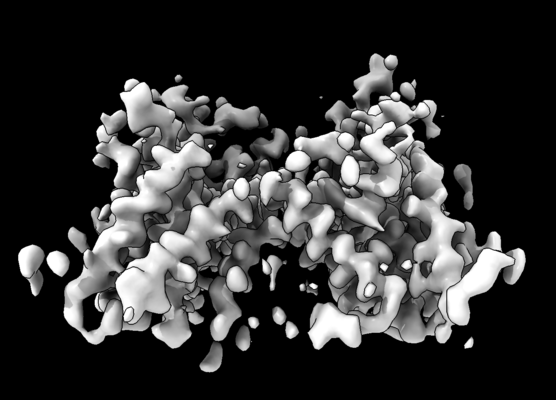
 Downloads & links
 emd_30508.png
 http://ftp.pdbj.org/pub/emdb/structures/EMD-30508


 Z (Sec.)
 Y (Row.)
 X (Col.)


 Sample components
 Processing
 Electron microscopy
 FIELD EMISSION GUN
