 ムービー
ムービー コントローラー


+ データを開く

- 基本情報
基本情報
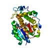
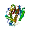
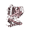
| 登録情報 | データベース: PDB / ID: 7cwj | ||||||
|---|---|---|---|---|---|---|---|
| タイトル | Root induced Secreted protein Tsp1 from Biocontrol fungi Trichoderma virens | ||||||
 要素 要素 | Root induced effector protein Tsp1 | ||||||
 キーワード キーワード | SIGNALING PROTEIN / Effector / ellicitor / bio-control agent | ||||||
| 機能・相同性 | effector-mediated perturbation of host innate immune response by symbiont / extracellular region / Effector TSP1 機能・相同性情報 機能・相同性情報 | ||||||
| 生物種 |  Hypocrea virens (菌類) Hypocrea virens (菌類) | ||||||
| 手法 |  X線回折 / シンクロトロン / 単波長異常分散 / 解像度: 1.61 Å X線回折 / シンクロトロン / 単波長異常分散 / 解像度: 1.61 Å | ||||||
 データ登録者 データ登録者 | Gupta, G.D. | ||||||
 引用 引用 | ジャーナル: Int.J.Biol.Macromol. / 年: 2021 タイトル: Structure-function analysis reveals Trichoderma virens Tsp1 to be a novel fungal effector protein modulating plant defence. 著者: Gupta, G.D. / Bansal, R. / Mistry, H. / Pandey, B. / Mukherjee, P.K. | ||||||
| 履歴 |
|
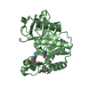
- 構造の表示
構造の表示
| 構造ビューア | 分子: MolmilJmol/JSmol |
|---|
- ダウンロードとリンク
-ダウンロード
| PDBx/mmCIF形式 | 7cwj.cif.gz | 298.6 KB | 表示 | PDBx/mmCIF形式 |
|---|---|---|---|---|
| PDB形式 | pdb7cwj.ent.gz | 247.1 KB | 表示 | PDB形式 |
| PDBx/mmJSON形式 | 7cwj.json.gz | ツリー表示 | PDBx/mmJSON形式 | |
| その他 |  その他のダウンロード その他のダウンロード |
-検証レポート
| アーカイブディレクトリ | https://data.pdbj.org/pub/pdb/validation_reports/cw/7cwjftp://data.pdbj.org/pub/pdb/validation_reports/cw/7cwj | HTTPS FTP |
|---|
-関連構造データ


-リンク
 PDBj
PDBj- 集合体
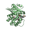
集合体
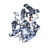
| 登録構造単位 | 
| ||||||||
|---|---|---|---|---|---|---|---|---|---|
| 1 | 
| ||||||||
| 2 | 
| ||||||||
| 単位格子 |
|
-要素
| #1: タンパク質 | 分子量: 14848.104 Da / 分子数: 4 / 由来タイプ: 組換発現 由来: (組換発現) Hypocrea virens (strain Gv29-8 / FGSC 10586) (菌類)株: Gv29-8 / FGSC 10586 / 遺伝子: TRIVIDRAFT_215947 / 発現宿主:  #2: 化合物 | ChemComp-GOL /   分子量: 92.094 Da / 分子数: 4 / 由来タイプ: 合成 / 式: C3H8O3 分子量: 92.094 Da / 分子数: 4 / 由来タイプ: 合成 / 式: C3H8O3#3: 水 | ChemComp-HOH / |  分子量: 18.015 Da / 分子数: 626 / 由来タイプ: 天然 / 式: H2O 分子量: 18.015 Da / 分子数: 626 / 由来タイプ: 天然 / 式: H2O研究の焦点であるリガンドがあるか | N | Has protein modification | Y | |
|---|
-実験情報
-実験
| 実験 | 手法: X線回折 / 使用した結晶の数: 1 |
|---|
- 試料調製
試料調製
| 結晶 | マシュー密度: 2.32 Å3/Da / 溶媒含有率: 47 % |
|---|---|
| 結晶化 | 温度: 289 K / 手法: 蒸気拡散法, ハンギングドロップ法 / pH: 6.5 / 詳細: 1.9 M sodium malonate pH 6.5, 5% glycerol |
-データ収集
| 回折 | 平均測定温度: 100 K / Serial crystal experiment: N | |||||||||||||||||||||||||||
|---|---|---|---|---|---|---|---|---|---|---|---|---|---|---|---|---|---|---|---|---|---|---|---|---|---|---|---|---|
| 放射光源 | 由来: シンクロトロン / サイト: RRCAT INDUS-2  / ビームライン: PX-BL21 / 波長: 0.9788 Å / ビームライン: PX-BL21 / 波長: 0.9788 Å | |||||||||||||||||||||||||||
| 検出器 | タイプ: RAYONIX MX340-HS / 検出器: CCD / 日付: 2016年10月13日 | |||||||||||||||||||||||||||
| 放射 | プロトコル: SINGLE WAVELENGTH / 単色(M)・ラウエ(L): M / 散乱光タイプ: x-ray | |||||||||||||||||||||||||||
| 放射波長 | 波長: 0.9788 Å / 相対比: 1 | |||||||||||||||||||||||||||
| 反射 | 解像度: 1.6→44.1 Å / Num. obs: 67496 / % possible obs: 96.2 % / 冗長度: 12.1 % / Biso Wilson estimate: 17.55 Å2 / CC1/2: 0.999 / Rmerge(I) obs: 0.067 / Rpim(I) all: 0.019 / Rrim(I) all: 0.07 / Net I/σ(I): 27.5 / Num. measured all: 818842 / Scaling rejects: 4 | |||||||||||||||||||||||||||
| 反射 シェル | Diffraction-ID: 1
|
-位相決定
| 位相決定 | 手法: 単波長異常分散 |
|---|
- 解析
解析
| ソフトウェア |
| ||||||||||||||||||||||||||||||||||||||||||||||||||||||||||||||||||||||||||||||||||||||||||||||||||||||||||||||||||||||||||||||||||||||||||||||||||||||
|---|---|---|---|---|---|---|---|---|---|---|---|---|---|---|---|---|---|---|---|---|---|---|---|---|---|---|---|---|---|---|---|---|---|---|---|---|---|---|---|---|---|---|---|---|---|---|---|---|---|---|---|---|---|---|---|---|---|---|---|---|---|---|---|---|---|---|---|---|---|---|---|---|---|---|---|---|---|---|---|---|---|---|---|---|---|---|---|---|---|---|---|---|---|---|---|---|---|---|---|---|---|---|---|---|---|---|---|---|---|---|---|---|---|---|---|---|---|---|---|---|---|---|---|---|---|---|---|---|---|---|---|---|---|---|---|---|---|---|---|---|---|---|---|---|---|---|---|---|---|---|---|
| 精密化 | 構造決定の手法: 単波長異常分散 / 解像度: 1.61→23.952 Å / SU ML: 0.17 / 交差検証法: THROUGHOUT / σ(F): 1.37 / 位相誤差: 19.5 / 立体化学のターゲット値: MLHL
| ||||||||||||||||||||||||||||||||||||||||||||||||||||||||||||||||||||||||||||||||||||||||||||||||||||||||||||||||||||||||||||||||||||||||||||||||||||||
| 溶媒の処理 | 減衰半径: 0.9 Å / VDWプローブ半径: 1.11 Å / 溶媒モデル: FLAT BULK SOLVENT MODEL | ||||||||||||||||||||||||||||||||||||||||||||||||||||||||||||||||||||||||||||||||||||||||||||||||||||||||||||||||||||||||||||||||||||||||||||||||||||||
| 原子変位パラメータ | Biso max: 80.42 Å2 / Biso mean: 23.802 Å2 / Biso min: 10.17 Å2 | ||||||||||||||||||||||||||||||||||||||||||||||||||||||||||||||||||||||||||||||||||||||||||||||||||||||||||||||||||||||||||||||||||||||||||||||||||||||
| 精密化ステップ | サイクル: final / 解像度: 1.61→23.952 Å
| ||||||||||||||||||||||||||||||||||||||||||||||||||||||||||||||||||||||||||||||||||||||||||||||||||||||||||||||||||||||||||||||||||||||||||||||||||||||
| 拘束条件 |
| ||||||||||||||||||||||||||||||||||||||||||||||||||||||||||||||||||||||||||||||||||||||||||||||||||||||||||||||||||||||||||||||||||||||||||||||||||||||
| LS精密化 シェル | Refine-ID: X-RAY DIFFRACTION / Rfactor Rfree error: 0
|
