 Movie
Movie Controller

[English] 日本語
 Yorodumi
- PDB-6ece: Vlm2 thioesterase domain with genetically encoded 2,3-diaminoprop... -
+ Open data

- Basic information
| Entry | Database: PDB / ID: 6ece | ||||||
|---|---|---|---|---|---|---|---|
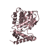
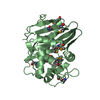
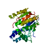
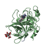
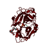
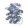
| Title | Vlm2 thioesterase domain with genetically encoded 2,3-diaminopropionic acid bound with a dodecadepsipeptide, space group H3 | ||||||
 Components Components |
| ||||||
 Keywords Keywords | HYDROLASE / thioesterase / thioesterase domain / NRPS / non-ribosomal peptide synthetase / nonribosomal peptide synthetase / valinomycin / depsipeptide / unnatural amino acid / 2 / 3-diaminopropionic acid | ||||||
| Function / homology |  Function and homology information Function and homology informationmonocarboxylic acid biosynthetic process / amino acid activation for nonribosomal peptide biosynthetic process / secondary metabolite biosynthetic process / lipid biosynthetic process / catalytic activity / phosphopantetheine binding / antibiotic biosynthetic process / cytoplasm Similarity search - Function | ||||||
| Biological species |  Streptomyces tsusimaensis (bacteria) Streptomyces tsusimaensis (bacteria)synthetic construct (others) | ||||||
| Method |  X-RAY DIFFRACTION / SYNCHROTRON / MOLECULAR REPLACEMENT / Resolution: 2 Å X-RAY DIFFRACTION / SYNCHROTRON / MOLECULAR REPLACEMENT / Resolution: 2 Å | ||||||
 Authors Authors | Alonzo, D.A. / Huguenin-Dezot, N. / Heberlig, G.W. / Mahesh, M. / Nguyen, D.P. / Dornan, M.H. / Boddy, C.N. / Chin, J.W. / Schmeing, T.M. | ||||||
| Funding support |  Canada, 1items Canada, 1items
| ||||||
 Citation Citation | Journal: Nature / Year: 2019 Title: Trapping biosynthetic acyl-enzyme intermediates with encoded 2,3-diaminopropionic acid. Authors: Huguenin-Dezot, N. / Alonzo, D.A. / Heberlig, G.W. / Mahesh, M. / Nguyen, D.P. / Dornan, M.H. / Boddy, C.N. / Schmeing, T.M. / Chin, J.W. | ||||||
| History |
|
- Structure visualization
| Structure viewer | Molecule: MolmilJmol/JSmol |
|---|
- Downloads & links
-Download
| PDBx/mmCIF format | 6ece.cif.gz | 330.5 KB | Display | PDBx/mmCIF format |
|---|---|---|---|---|
| PDB format | pdb6ece.ent.gz | 223.9 KB | Display | PDB format |
| PDBx/mmJSON format | 6ece.json.gz | Tree view | PDBx/mmJSON format | |
| Others |  Other downloads Other downloads |
-Validation report
| Arichive directory | https://data.pdbj.org/pub/pdb/validation_reports/ec/6eceftp://data.pdbj.org/pub/pdb/validation_reports/ec/6ece | HTTPS FTP |
|---|
-Related structure data
| Related structure data |  6ecbSC  6eccC 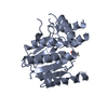 6ecdC 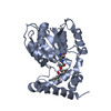 6ecfC S: Starting model for refinement C: citing same article ( |
|---|---|
| Similar structure data |

-Links
 PDBj
PDBj


- Assembly
| Deposited unit | 
| ||||||||||||
|---|---|---|---|---|---|---|---|---|---|---|---|---|---|
| 1 | 
| ||||||||||||
| 2 | 
| ||||||||||||
| Unit cell |
| ||||||||||||
| Components on special symmetry positions |
|
-Components
| #1: Protein | Mass: 33095.512 Da / Num. of mol.: 2 / Fragment: thioesterase domain (UNP residues 2368-2655) Source method: isolated from a genetically manipulated source Source: (gene. exp.) Streptomyces tsusimaensis (bacteria) / Gene: vlm2 / Production host: References: UniProt: Q1PSF3, Hydrolases; Acting on ester bonds; Thioester hydrolases #2: Protein/peptide | Mass: 1129.336 Da / Num. of mol.: 2 / Source method: obtained synthetically / Source: (synth.) synthetic construct (others) #3: Water | ChemComp-HOH / |  Mass: 18.015 Da / Num. of mol.: 89 / Source method: isolated from a natural source / Formula: H2O Mass: 18.015 Da / Num. of mol.: 89 / Source method: isolated from a natural source / Formula: H2O |
|---|
-Experimental details
-Experiment
| Experiment | Method: X-RAY DIFFRACTION / Number of used crystals: 1 |
|---|
- Sample preparation
| Crystal | Density Matthews: 2.04 Å3/Da / Density % sol: 39.84 % |
|---|---|
| Crystal grow | Temperature: 295 K / Method: vapor diffusion, hanging drop / pH: 8 Details: 1.3 M DL-malic acid, pH 8.1, 25 mM HEPES, pH 8.0, 100 mM sodium chloride, 0.2 mM TCEP |
-Data collection
| Diffraction | Mean temperature: 100 K |
|---|---|
| Diffraction source | Source: SYNCHROTRON / Site: CLSI / Beamline: 08ID-1 / Wavelength: 0.9794 Å |
| Detector | Type: DECTRIS PILATUS3 S 6M / Detector: PIXEL / Date: Feb 15, 2018 |
| Radiation | Monochromator: double crystal Si(111) / Protocol: SINGLE WAVELENGTH / Monochromatic (M) / Laue (L): M / Scattering type: x-ray |
| Radiation wavelength | Wavelength: 0.9794 Å / Relative weight: 1 |
| Reflection | Resolution: 2→64.59 Å / Num. obs: 35685 / % possible obs: 100 % / Redundancy: 5 % / Biso Wilson estimate: 39.62 Å2 / CC1/2: 0.998 / Rmerge(I) obs: 0.079 / Rpim(I) all: 0.053 / Rrim(I) all: 0.088 / Χ2: 0.89 / Net I/σ(I): 9.8 |
| Reflection shell | Resolution: 2→2.05 Å / Num. measured obs: 13233 / Num. unique obs: 2682 / CC1/2: 0.382 |
- Processing
| Software |
| ||||||||||||||||||||||||||||||||||||||||||||||||||||||||||||||||||||||||||||||||||||||||||||||||||
|---|---|---|---|---|---|---|---|---|---|---|---|---|---|---|---|---|---|---|---|---|---|---|---|---|---|---|---|---|---|---|---|---|---|---|---|---|---|---|---|---|---|---|---|---|---|---|---|---|---|---|---|---|---|---|---|---|---|---|---|---|---|---|---|---|---|---|---|---|---|---|---|---|---|---|---|---|---|---|---|---|---|---|---|---|---|---|---|---|---|---|---|---|---|---|---|---|---|---|---|
| Refinement | Method to determine structure: MOLECULAR REPLACEMENT Starting model: PDB entry 6ECB Resolution: 2→64.59 Å / SU ML: 0.2718 / Cross valid method: FREE R-VALUE / σ(F): 2.09 / Phase error: 26.854
| ||||||||||||||||||||||||||||||||||||||||||||||||||||||||||||||||||||||||||||||||||||||||||||||||||
| Solvent computation | Shrinkage radii: 0.9 Å / VDW probe radii: 1.11 Å | ||||||||||||||||||||||||||||||||||||||||||||||||||||||||||||||||||||||||||||||||||||||||||||||||||
| Displacement parameters | Biso mean: 52.5 Å2 | ||||||||||||||||||||||||||||||||||||||||||||||||||||||||||||||||||||||||||||||||||||||||||||||||||
| Refinement step | Cycle: LAST / Resolution: 2→64.59 Å
| ||||||||||||||||||||||||||||||||||||||||||||||||||||||||||||||||||||||||||||||||||||||||||||||||||
| Refine LS restraints |
| ||||||||||||||||||||||||||||||||||||||||||||||||||||||||||||||||||||||||||||||||||||||||||||||||||
| LS refinement shell |
| ||||||||||||||||||||||||||||||||||||||||||||||||||||||||||||||||||||||||||||||||||||||||||||||||||
| Refinement TLS params. | Method: refined / Origin x: 15.5882246133 Å / Origin y: -22.9006581781 Å / Origin z: -2.6962886655 Å
| ||||||||||||||||||||||||||||||||||||||||||||||||||||||||||||||||||||||||||||||||||||||||||||||||||
| Refinement TLS group | Selection details: all |
