 Movie
Movie Controller

[English] 日本語
 Yorodumi
- PDB-7cmq: Crystal Structure of Bacillus sp. TB-90 Urate Oxidase Improved by... -
+ Open data

- Basic information
| Entry | Database: PDB / ID: 7cmq | |||||||||
|---|---|---|---|---|---|---|---|---|---|---|
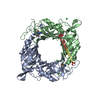
| Title | Crystal Structure of Bacillus sp. TB-90 Urate Oxidase Improved by Humidity Control at 88% RH. | |||||||||
 Components Components | Uric acid degradation bifunctional protein | |||||||||
 Keywords Keywords | OXIDOREDUCTASE / protein engineering / enzyme / loop flexibility / entropy of activation | |||||||||
| Function / homology |  Function and homology information Function and homology information2-oxo-4-hydroxy-4-carboxy-5-ureidoimidazoline decarboxylase / 2-oxo-4-hydroxy-4-carboxy-5-ureidoimidazoline decarboxylase activity / factor-independent urate hydroxylase / urate oxidase activity / urate catabolic process / allantoin metabolic process / purine nucleobase metabolic process Similarity search - Function | |||||||||
| Biological species |  | |||||||||
| Method |  X-RAY DIFFRACTION / SYNCHROTRON / MOLECULAR REPLACEMENT / Resolution: 1.65 Å X-RAY DIFFRACTION / SYNCHROTRON / MOLECULAR REPLACEMENT / Resolution: 1.65 Å | |||||||||
 Authors Authors | Hibi, T. / Itoh, T. / Nishiya, Y. | |||||||||
 Citation Citation | Journal: To be published Title: Flexibility of a Distal Interface Loop Modulates Water Network in the Active Site of Bacillus sp. TB-90 Urate Oxidase Authors: Hibi, T. / Itoh, T. / Nishiya, Y. #1: Journal: Biochemistry / Year: 2016Title: Hyperstabilization of Tetrameric Bacillus sp. TB-90 Urate Oxidase by Introducing Disulfide Bonds through Structural Plasticity. Authors: Hibi, T. / Kume, A. / Kawamura, A. / Itoh, T. / Fukada, H. / Nishiya, Y. #2: Journal: Biochemistry / Year: 2014Title: Intersubunit salt bridges with a sulfate anion control subunit dissociation and thermal stabilization of Bacillus sp. TB-90 urate oxidase. Authors: Hibi, T. / Hayashi, Y. / Fukada, H. / Itoh, T. / Nago, T. / Nishiya, Y. | |||||||||
| History |
|
- Structure visualization
| Structure viewer | Molecule: MolmilJmol/JSmol |
|---|
- Downloads & links
-Download
| PDBx/mmCIF format | 7cmq.cif.gz | 445.5 KB | Display | PDBx/mmCIF format |
|---|---|---|---|---|
| PDB format | pdb7cmq.ent.gz | 306.7 KB | Display | PDB format |
| PDBx/mmJSON format | 7cmq.json.gz | Tree view | PDBx/mmJSON format | |
| Others |  Other downloads Other downloads |
-Validation report
| Arichive directory | https://data.pdbj.org/pub/pdb/validation_reports/cm/7cmqftp://data.pdbj.org/pub/pdb/validation_reports/cm/7cmq | HTTPS FTP |
|---|
-Related structure data
| Related structure data |  5yjaC  5z27C  5z2bC  7cmnC  3wlvS S: Starting model for refinement C: citing same article ( |
|---|---|
| Similar structure data |


-Links
 PDBj
PDBj


- Assembly
| Deposited unit | 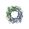
| ||||||||||||
|---|---|---|---|---|---|---|---|---|---|---|---|---|---|
| 1 | 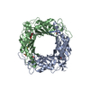
| ||||||||||||
| Unit cell |
|
-Components
-Protein , 1 types, 2 molecules AB
| #1: Protein | Mass: 35823.301 Da / Num. of mol.: 2 Source method: isolated from a genetically manipulated source Source: (gene. exp.) References: UniProt: Q45697, factor-independent urate hydroxylase |
|---|
-Non-polymers , 5 types, 396 molecules 


| #2: Chemical |  Mass: 153.099 Da / Num. of mol.: 2 / Source method: obtained synthetically / Formula: C4H3N5O2 / Feature type: SUBJECT OF INVESTIGATION Mass: 153.099 Da / Num. of mol.: 2 / Source method: obtained synthetically / Formula: C4H3N5O2 / Feature type: SUBJECT OF INVESTIGATION#3: Chemical |  Mass: 31.999 Da / Num. of mol.: 2 / Source method: obtained synthetically / Formula: O2 / Feature type: SUBJECT OF INVESTIGATION Mass: 31.999 Da / Num. of mol.: 2 / Source method: obtained synthetically / Formula: O2 / Feature type: SUBJECT OF INVESTIGATION#4: Chemical | ChemComp-SO4 / |  Mass: 96.063 Da / Num. of mol.: 1 / Source method: obtained synthetically / Formula: SO4 Mass: 96.063 Da / Num. of mol.: 1 / Source method: obtained synthetically / Formula: SO4#5: Chemical | ChemComp-EDO /  Mass: 62.068 Da / Num. of mol.: 5 / Source method: obtained synthetically / Formula: C2H6O2 Mass: 62.068 Da / Num. of mol.: 5 / Source method: obtained synthetically / Formula: C2H6O2#6: Water | ChemComp-HOH / | Mass: 18.015 Da / Num. of mol.: 386 / Source method: isolated from a natural source / Formula: H2O |
|---|
-Details
| Has ligand of interest | Y |
|---|
-Experimental details
-Experiment
| Experiment | Method: X-RAY DIFFRACTION / Number of used crystals: 1 |
|---|
- Sample preparation
| Crystal | Density Matthews: 2.43 Å3/Da / Density % sol: 49.5 % |
|---|---|
| Crystal grow | Temperature: 293 K / Method: vapor diffusion, hanging drop / pH: 8.5 Details: 16% PEG 8000, 0.1 M Tris-HCl, 0.08 M K2SO4, 2 mM 8-azaxthantine |
-Data collection
| Diffraction | Mean temperature: 100 K / Serial crystal experiment: N |
|---|---|
| Diffraction source | Source: SYNCHROTRON / Site: SPring-8  / Beamline: BL38B1 / Wavelength: 1 Å / Beamline: BL38B1 / Wavelength: 1 Å |
| Detector | Type: ADSC QUANTUM 315r / Detector: CCD / Date: Jul 19, 2017 |
| Radiation | Protocol: SINGLE WAVELENGTH / Monochromatic (M) / Laue (L): M / Scattering type: x-ray |
| Radiation wavelength | Wavelength: 1 Å / Relative weight: 1 |
| Reflection | Resolution: 1.65→46.1 Å / Num. obs: 82336 / % possible obs: 99.4 % / Redundancy: 3.8 % / Biso Wilson estimate: 24.36 Å2 / CC1/2: 0.998 / Rmerge(I) obs: 0.054 / Rrim(I) all: 0.063 / Net I/σ(I): 13.96 |
| Reflection shell | Resolution: 1.65→1.75 Å / Redundancy: 3.7 % / Rmerge(I) obs: 0.673 / Mean I/σ(I) obs: 2.12 / Num. unique obs: 25541 / CC1/2: 0.723 / Rrim(I) all: 0.788 / % possible all: 98.8 |
- Processing
| Software |
| ||||||||||||||||||||||||||||||||||||||||||||||||||||||||||||||||||||||||||||||||||||||||||||||||||||||||||||||||||||||||||||||||||||||||||||||||||||||||||||||||||||||||||||||||||||||||||||||||||||||||||||||||||
|---|---|---|---|---|---|---|---|---|---|---|---|---|---|---|---|---|---|---|---|---|---|---|---|---|---|---|---|---|---|---|---|---|---|---|---|---|---|---|---|---|---|---|---|---|---|---|---|---|---|---|---|---|---|---|---|---|---|---|---|---|---|---|---|---|---|---|---|---|---|---|---|---|---|---|---|---|---|---|---|---|---|---|---|---|---|---|---|---|---|---|---|---|---|---|---|---|---|---|---|---|---|---|---|---|---|---|---|---|---|---|---|---|---|---|---|---|---|---|---|---|---|---|---|---|---|---|---|---|---|---|---|---|---|---|---|---|---|---|---|---|---|---|---|---|---|---|---|---|---|---|---|---|---|---|---|---|---|---|---|---|---|---|---|---|---|---|---|---|---|---|---|---|---|---|---|---|---|---|---|---|---|---|---|---|---|---|---|---|---|---|---|---|---|---|---|---|---|---|---|---|---|---|---|---|---|---|---|---|---|---|---|
| Refinement | Method to determine structure: MOLECULAR REPLACEMENT Starting model: 3WLV Resolution: 1.65→46.1 Å / SU ML: 0.1717 / Cross valid method: FREE R-VALUE / σ(F): 1.34 / Phase error: 19.0511 Stereochemistry target values: GeoStd + Monomer Library + CDL v1.2
| ||||||||||||||||||||||||||||||||||||||||||||||||||||||||||||||||||||||||||||||||||||||||||||||||||||||||||||||||||||||||||||||||||||||||||||||||||||||||||||||||||||||||||||||||||||||||||||||||||||||||||||||||||
| Solvent computation | Shrinkage radii: 0.9 Å / VDW probe radii: 1.11 Å / Solvent model: FLAT BULK SOLVENT MODEL | ||||||||||||||||||||||||||||||||||||||||||||||||||||||||||||||||||||||||||||||||||||||||||||||||||||||||||||||||||||||||||||||||||||||||||||||||||||||||||||||||||||||||||||||||||||||||||||||||||||||||||||||||||
| Displacement parameters | Biso mean: 29.69 Å2 | ||||||||||||||||||||||||||||||||||||||||||||||||||||||||||||||||||||||||||||||||||||||||||||||||||||||||||||||||||||||||||||||||||||||||||||||||||||||||||||||||||||||||||||||||||||||||||||||||||||||||||||||||||
| Refine analyze | Luzzati coordinate error obs: 0.219 Å | ||||||||||||||||||||||||||||||||||||||||||||||||||||||||||||||||||||||||||||||||||||||||||||||||||||||||||||||||||||||||||||||||||||||||||||||||||||||||||||||||||||||||||||||||||||||||||||||||||||||||||||||||||
| Refinement step | Cycle: LAST / Resolution: 1.65→46.1 Å
| ||||||||||||||||||||||||||||||||||||||||||||||||||||||||||||||||||||||||||||||||||||||||||||||||||||||||||||||||||||||||||||||||||||||||||||||||||||||||||||||||||||||||||||||||||||||||||||||||||||||||||||||||||
| Refine LS restraints |
| ||||||||||||||||||||||||||||||||||||||||||||||||||||||||||||||||||||||||||||||||||||||||||||||||||||||||||||||||||||||||||||||||||||||||||||||||||||||||||||||||||||||||||||||||||||||||||||||||||||||||||||||||||
| LS refinement shell |
| ||||||||||||||||||||||||||||||||||||||||||||||||||||||||||||||||||||||||||||||||||||||||||||||||||||||||||||||||||||||||||||||||||||||||||||||||||||||||||||||||||||||||||||||||||||||||||||||||||||||||||||||||||
| Refinement TLS params. | Method: refined / Origin x: 13.9650704308 Å / Origin y: 4.19013357373 Å / Origin z: 38.4381304565 Å
| ||||||||||||||||||||||||||||||||||||||||||||||||||||||||||||||||||||||||||||||||||||||||||||||||||||||||||||||||||||||||||||||||||||||||||||||||||||||||||||||||||||||||||||||||||||||||||||||||||||||||||||||||||
| Refinement TLS group | Selection details: all |
