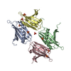
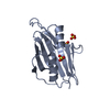
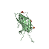
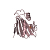
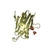
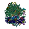
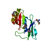
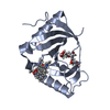
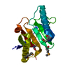
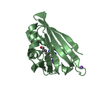
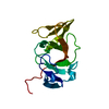
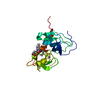
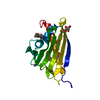
Journal: Nat Commun / Year: 2021 Title: Structural and molecular basis for Cardiovirus 2A protein as a viral gene expression switch. Authors: Chris H Hill / Lukas Pekarek / Sawsan Napthine / Anuja Kibe / Andrew E Firth / Stephen C Graham / Neva Caliskan / Ian Brierley / Abstract: Programmed -1 ribosomal frameshifting (PRF) in cardioviruses is activated by the 2A protein, a multi-functional virulence factor that also inhibits cap-dependent translational initiation. Here we ...Programmed -1 ribosomal frameshifting (PRF) in cardioviruses is activated by the 2A protein, a multi-functional virulence factor that also inhibits cap-dependent translational initiation. Here we present the X-ray crystal structure of 2A and show that it selectively binds to a pseudoknot-like conformation of the PRF stimulatory RNA element in the viral genome. Using optical tweezers, we demonstrate that 2A stabilises this RNA element, likely explaining the increase in PRF efficiency in the presence of 2A. Next, we demonstrate a strong interaction between 2A and the small ribosomal subunit and present a cryo-EM structure of 2A bound to initiated 70S ribosomes. Multiple copies of 2A bind to the 16S rRNA where they may compete for binding with initiation and elongation factors. Together, these results define the structural basis for RNA recognition by 2A, show how 2A-mediated stabilisation of an RNA pseudoknot promotes PRF, and reveal how 2A accumulation may shut down translation during virus infection.
Evidence: light scattering, In solution, protein is monomeric as informed by size-exclusion chromatography coupled to multi-angle light scattering (SEC-MALS)
 Movie
Movie Controller


 Open data
 Basic information
 Components
 Keywords
 Function and homology information
 Mengo encephalomyocarditis virus
 X-RAY DIFFRACTION /
  Authors
 United Kingdom, European Union, 3items
  Citation
 Structure visualization
 Downloads & links
 Other downloads
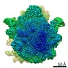


 PDBj
PDBj

 Assembly