Movie
Movie Controller

[English] 日本語
 Yorodumi
- PDB-7nwt: Initiated 70S ribosome in complex with 2A protein from encephalom... -
+ Open data

- Basic information
| Entry | Database: PDB / ID: 7nwt | ||||||||||||||||||||||||||||||||||||||||||||||||||||||
|---|---|---|---|---|---|---|---|---|---|---|---|---|---|---|---|---|---|---|---|---|---|---|---|---|---|---|---|---|---|---|---|---|---|---|---|---|---|---|---|---|---|---|---|---|---|---|---|---|---|---|---|---|---|---|---|
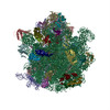
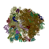
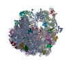
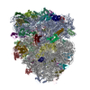
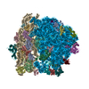
| Title | Initiated 70S ribosome in complex with 2A protein from encephalomyocarditis virus (EMCV) | ||||||||||||||||||||||||||||||||||||||||||||||||||||||
 Components Components |
| ||||||||||||||||||||||||||||||||||||||||||||||||||||||
 Keywords Keywords | RIBOSOME / 70S ribosome / initiation complex / EMCV / cardiovirus / 2A / picornavirus / frameshifting / PRF / RNA-binding protein / protein-mediated frameshifting / ribosome-binding protein / beta-shell / viral protein | ||||||||||||||||||||||||||||||||||||||||||||||||||||||
| Function / homology |  Function and homology information Function and homology informationpositive stranded viral RNA replication / host cell nucleolus / stringent response / symbiont-mediated suppression of host cytoplasmic pattern recognition receptor signaling pathway via inhibition of RIG-I activity / misfolded RNA binding / Group I intron splicing / RNA folding / translational termination / transcriptional attenuation / endoribonuclease inhibitor activity ...positive stranded viral RNA replication / host cell nucleolus / stringent response / symbiont-mediated suppression of host cytoplasmic pattern recognition receptor signaling pathway via inhibition of RIG-I activity / misfolded RNA binding / Group I intron splicing / RNA folding / translational termination / transcriptional attenuation / endoribonuclease inhibitor activity / positive regulation of ribosome biogenesis / RNA-binding transcription regulator activity / negative regulation of cytoplasmic translation / DnaA-L2 complex / translation repressor activity / negative regulation of DNA-templated DNA replication initiation / mRNA regulatory element binding translation repressor activity / positive regulation of RNA splicing / response to reactive oxygen species / cytosolic ribosome assembly / ribosome assembly / assembly of large subunit precursor of preribosome / picornain 3C / regulation of cell growth / T=pseudo3 icosahedral viral capsid / DNA-templated transcription termination / response to radiation / host cell cytoplasmic vesicle membrane / maintenance of translational fidelity / mRNA 5'-UTR binding / large ribosomal subunit / transferase activity / ribosomal small subunit assembly / channel activity / ribosome binding / ribosome biogenesis / ribosomal small subunit biogenesis / 5S rRNA binding / ribosomal large subunit assembly / small ribosomal subunit / small ribosomal subunit rRNA binding / cytosolic small ribosomal subunit / large ribosomal subunit rRNA binding / monoatomic ion transmembrane transport / cytosolic large ribosomal subunit / cytoplasmic translation / tRNA binding / RNA helicase activity / negative regulation of translation / rRNA binding / RNA helicase / structural constituent of ribosome / ribosome / translation / symbiont-mediated suppression of host gene expression / ribonucleoprotein complex / viral translational frameshifting / symbiont-mediated activation of host autophagy / RNA-directed RNA polymerase / cysteine-type endopeptidase activity / response to antibiotic / negative regulation of DNA-templated transcription / RNA-directed RNA polymerase activity / mRNA binding / symbiont entry into host cell / virion attachment to host cell / structural molecule activity / ATP hydrolysis activity / DNA-templated transcription / proteolysis / DNA binding / RNA binding / zinc ion binding / ATP binding / membrane / metal ion binding / cytosol / cytoplasm Similarity search - Function | ||||||||||||||||||||||||||||||||||||||||||||||||||||||
| Biological species |  Mengo encephalomyocarditis virus Mengo encephalomyocarditis virus Encephalomyocarditis virus Encephalomyocarditis virus | ||||||||||||||||||||||||||||||||||||||||||||||||||||||
| Method | ELECTRON MICROSCOPY / single particle reconstruction / cryo EM / Resolution: 2.66 Å | ||||||||||||||||||||||||||||||||||||||||||||||||||||||
 Authors Authors | Hill, C.H. / Napthine, S. / Pekarek, L. / Kibe, A. / Firth, A.E. / Graham, S.C. / Caliskan, N. / Brierley, I. | ||||||||||||||||||||||||||||||||||||||||||||||||||||||
| Funding support |  United Kingdom, European Union, 3items United Kingdom, European Union, 3items
| ||||||||||||||||||||||||||||||||||||||||||||||||||||||
 Citation Citation | Journal: Nat Commun / Year: 2021 Title: Structural and molecular basis for Cardiovirus 2A protein as a viral gene expression switch. Authors: Chris H Hill / Lukas Pekarek / Sawsan Napthine / Anuja Kibe / Andrew E Firth / Stephen C Graham / Neva Caliskan / Ian Brierley /  Abstract: Programmed -1 ribosomal frameshifting (PRF) in cardioviruses is activated by the 2A protein, a multi-functional virulence factor that also inhibits cap-dependent translational initiation. Here we ...Programmed -1 ribosomal frameshifting (PRF) in cardioviruses is activated by the 2A protein, a multi-functional virulence factor that also inhibits cap-dependent translational initiation. Here we present the X-ray crystal structure of 2A and show that it selectively binds to a pseudoknot-like conformation of the PRF stimulatory RNA element in the viral genome. Using optical tweezers, we demonstrate that 2A stabilises this RNA element, likely explaining the increase in PRF efficiency in the presence of 2A. Next, we demonstrate a strong interaction between 2A and the small ribosomal subunit and present a cryo-EM structure of 2A bound to initiated 70S ribosomes. Multiple copies of 2A bind to the 16S rRNA where they may compete for binding with initiation and elongation factors. Together, these results define the structural basis for RNA recognition by 2A, show how 2A-mediated stabilisation of an RNA pseudoknot promotes PRF, and reveal how 2A accumulation may shut down translation during virus infection. | ||||||||||||||||||||||||||||||||||||||||||||||||||||||
| History |
|
- Structure visualization
| Movie |
Movie viewer |
|---|---|
| Structure viewer | Molecule: MolmilJmol/JSmol |
- Downloads & links
-Download
| PDBx/mmCIF format | 7nwt.cif.gz | 3.9 MB | Display | PDBx/mmCIF format |
|---|---|---|---|---|
| PDB format | pdb7nwt.ent.gz | Display | PDB format | |
| PDBx/mmJSON format | 7nwt.json.gz | Tree view | PDBx/mmJSON format | |
| Others |  Other downloads Other downloads |
-Validation report
| Arichive directory | https://data.pdbj.org/pub/pdb/validation_reports/nw/7nwtftp://data.pdbj.org/pub/pdb/validation_reports/nw/7nwt | HTTPS FTP |
|---|
-Related structure data
| Related structure data | 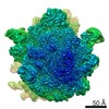 12635MC  7bnyC C: citing same article ( M: map data used to model this data |
|---|---|
| Similar structure data |


-Links
 PDBj
PDBj


- Assembly
| Deposited unit | 
|
|---|---|
| 1 |
|
-Components
+50S ribosomal protein ... , 31 types, 31 molecules BCDEFGHIJKLMNOPQRSTUVWXYZabcdef
-RNA chain , 5 types, 5 molecules 1235XX
| #26: RNA chain | Mass: 941832.625 Da / Num. of mol.: 1 / Source method: isolated from a natural source / Source: (natural) |
|---|---|
| #27: RNA chain | Mass: 497405.969 Da / Num. of mol.: 1 / Source method: isolated from a natural source / Source: (natural) |
| #28: RNA chain | Mass: 38790.090 Da / Num. of mol.: 1 / Source method: isolated from a natural source / Source: (natural) |
| #29: RNA chain | Mass: 24806.922 Da / Num. of mol.: 1 / Source method: isolated from a natural source / Source: (natural) |
| #56: RNA chain | Mass: 37684.395 Da / Num. of mol.: 1 / Source method: obtained synthetically / Details: in vitro transcription / Source: (synth.) Encephalomyocarditis virus |
-30S ribosomal protein ... , 20 types, 20 molecules ghijklmnopqrstuvwxyz
| #36: Protein | Mass: 26781.670 Da / Num. of mol.: 1 / Source method: isolated from a natural source / Source: (natural) |
|---|---|
| #37: Protein | Mass: 26031.316 Da / Num. of mol.: 1 / Source method: isolated from a natural source / Source: (natural) |
| #38: Protein | Mass: 23514.199 Da / Num. of mol.: 1 / Source method: isolated from a natural source / Source: (natural) |
| #39: Protein | Mass: 17629.398 Da / Num. of mol.: 1 / Source method: isolated from a natural source / Source: (natural) |
| #40: Protein | Mass: 15727.512 Da / Num. of mol.: 1 / Source method: isolated from a natural source / Source: (natural) |
| #41: Protein | Mass: 20055.156 Da / Num. of mol.: 1 / Source method: isolated from a natural source / Source: (natural) |
| #42: Protein | Mass: 14146.557 Da / Num. of mol.: 1 / Source method: isolated from a natural source / Source: (natural) |
| #43: Protein | Mass: 14886.270 Da / Num. of mol.: 1 / Source method: isolated from a natural source / Source: (natural) |
| #44: Protein | Mass: 11755.597 Da / Num. of mol.: 1 / Source method: isolated from a natural source / Source: (natural) |
| #45: Protein | Mass: 13870.975 Da / Num. of mol.: 1 / Source method: isolated from a natural source / Source: (natural) |
| #46: Protein | Mass: 13814.249 Da / Num. of mol.: 1 / Source method: isolated from a natural source / Source: (natural) |
| #47: Protein | Mass: 13128.467 Da / Num. of mol.: 1 / Source method: isolated from a natural source / Source: (natural) |
| #48: Protein | Mass: 11606.560 Da / Num. of mol.: 1 / Source method: isolated from a natural source / Source: (natural) |
| #49: Protein | Mass: 10290.816 Da / Num. of mol.: 1 / Source method: isolated from a natural source / Source: (natural) |
| #50: Protein | Mass: 9207.572 Da / Num. of mol.: 1 / Source method: isolated from a natural source / Source: (natural) |
| #51: Protein | Mass: 9724.491 Da / Num. of mol.: 1 / Source method: isolated from a natural source / Source: (natural) |
| #52: Protein | Mass: 9005.472 Da / Num. of mol.: 1 / Source method: isolated from a natural source / Source: (natural) |
| #53: Protein | Mass: 10455.355 Da / Num. of mol.: 1 / Source method: isolated from a natural source / Source: (natural) |
| #54: Protein | Mass: 9708.464 Da / Num. of mol.: 1 / Source method: isolated from a natural source / Source: (natural) |
| #55: Protein | Mass: 8524.039 Da / Num. of mol.: 1 / Source method: isolated from a natural source / Source: (natural) |
-Protein , 1 types, 3 molecules AABBCC
| #57: Protein | Mass: 17831.182 Da / Num. of mol.: 3 Source method: isolated from a genetically manipulated source Source: (gene. exp.) Mengo encephalomyocarditis virus / Production host: |
|---|
-Non-polymers , 4 types, 442 molecules 


| #58: Chemical | ChemComp-MG /  Mass: 24.305 Da / Num. of mol.: 437 / Source method: obtained synthetically / Formula: Mg Mass: 24.305 Da / Num. of mol.: 437 / Source method: obtained synthetically / Formula: Mg#59: Chemical | ChemComp-FME / |  Type: L-peptide linking / Mass: 177.221 Da / Num. of mol.: 1 / Source method: obtained synthetically / Formula: C6H11NO3S Type: L-peptide linking / Mass: 177.221 Da / Num. of mol.: 1 / Source method: obtained synthetically / Formula: C6H11NO3S#60: Chemical |  Mass: 65.409 Da / Num. of mol.: 2 / Source method: obtained synthetically / Formula: Zn Mass: 65.409 Da / Num. of mol.: 2 / Source method: obtained synthetically / Formula: Zn#61: Water | ChemComp-HOH / | Mass: 18.015 Da / Num. of mol.: 2 / Source method: isolated from a natural source / Formula: H2O |
|---|
-Details
| Has protein modification | Y |
|---|
-Experimental details
-Experiment
| Experiment | Method: ELECTRON MICROSCOPY |
|---|---|
| EM experiment | Aggregation state: PARTICLE / 3D reconstruction method: single particle reconstruction |
- Sample preparation
| Component | Name: Initiated 70S ribosome in complex with 2A protein from encephalomyocarditis virus (EMCV) Type: RIBOSOME Details: 70S ribosome subunits and initiator fMet-tRNA purified from E.coli. mRNA template was generated by in vitro transcription EMCV 2A protein was recombinantly expressed in E.coli Entity ID: #1-#28, #30-#57 / Source: MULTIPLE SOURCES | ||||||||||||||||||||||||||||||||
|---|---|---|---|---|---|---|---|---|---|---|---|---|---|---|---|---|---|---|---|---|---|---|---|---|---|---|---|---|---|---|---|---|---|
| Molecular weight | Experimental value: NO | ||||||||||||||||||||||||||||||||
| Buffer solution | pH: 7.5 Details: Initiated 70S ribosomes in 50 mM Tris-HCl pH 7.5, 70 mM NH4Cl, 30 mM KCl, 7 mM MgCl2 were diluted tenfold into 20 mM HEPES pH 7.5, 100 mM potassium acetate, 1.5 mM MgCl2, 2.0 mM DTT. 2A ...Details: Initiated 70S ribosomes in 50 mM Tris-HCl pH 7.5, 70 mM NH4Cl, 30 mM KCl, 7 mM MgCl2 were diluted tenfold into 20 mM HEPES pH 7.5, 100 mM potassium acetate, 1.5 mM MgCl2, 2.0 mM DTT. 2A protein was dialysed (3K MWCO, 277K, 16 h) into the same buffer. Crosslinking reactions of 50 microliters comprising 75 nM ribosomes, 3.0 micromolar 2A and 2.0 mM bis(sulfosuccinimidyl)suberate (BS3) were performed on ice (30 min) immediately prior to grid preparation. | ||||||||||||||||||||||||||||||||
| Buffer component |
| ||||||||||||||||||||||||||||||||
| Specimen | Embedding applied: NO / Shadowing applied: NO / Staining applied: NO / Vitrification applied: YES Details: 75 nM ribosomes, 3.0 micromolar 2A crosslinked with 2.0 mM bis(sulfosuccinimidyl)suberate (BS3) | ||||||||||||||||||||||||||||||||
| Specimen support | Grid material: COPPER / Grid mesh size: 400 divisions/in. / Grid type: Quantifoil R2/2 | ||||||||||||||||||||||||||||||||
| Vitrification | Instrument: FEI VITROBOT MARK IV / Cryogen name: ETHANE / Humidity: 100 % / Chamber temperature: 277 K Details: Quantifoil R 2/2 400-mesh copper supports were coated with an additional ~ 60 angstrom layer of amorphous, evaporated carbon by flotation and thoroughly dried before use. Grids were made ...Details: Quantifoil R 2/2 400-mesh copper supports were coated with an additional ~ 60 angstrom layer of amorphous, evaporated carbon by flotation and thoroughly dried before use. Grids were made hydrophilic by glow-discharge in air for 30 s. Three microliters of crosslinking reaction was applied to grids which were then blotted for 4.5 s and vitrified by plunging into liquid ethane using a Vitrobot MK IV (FEI) at 277K, 100% relative humidity. |
- Electron microscopy imaging
| Experimental equipment |  Model: Titan Krios / Image courtesy: FEI Company |
|---|---|
| Microscopy | Model: FEI TITAN KRIOS |
| Electron gun | Electron source:  FIELD EMISSION GUN / Accelerating voltage: 300 kV / Illumination mode: FLOOD BEAM FIELD EMISSION GUN / Accelerating voltage: 300 kV / Illumination mode: FLOOD BEAM |
| Electron lens | Mode: BRIGHT FIELD / Nominal magnification: 75000 X / Nominal defocus max: 3000 nm / Nominal defocus min: 1200 nm / Cs: 2.7 mm / C2 aperture diameter: 100 µm / Alignment procedure: COMA FREE |
| Specimen holder | Cryogen: NITROGEN / Specimen holder model: FEI TITAN KRIOS AUTOGRID HOLDER |
| Image recording | Average exposure time: 0.59 sec. / Electron dose: 54.4 e/Å2 / Detector mode: INTEGRATING / Film or detector model: FEI FALCON III (4k x 4k) / Num. of grids imaged: 1 / Num. of real images: 5730 Details: Images were collected as 23-frame movies. Autofocus every 10 micrometres, no drift measurement, 7 sec delay after stage shift and 3 sec delay after image shift |
- Processing
| Software |
| ||||||||||||||||||||||||||||||||||||||||||||||||||||||||||||
|---|---|---|---|---|---|---|---|---|---|---|---|---|---|---|---|---|---|---|---|---|---|---|---|---|---|---|---|---|---|---|---|---|---|---|---|---|---|---|---|---|---|---|---|---|---|---|---|---|---|---|---|---|---|---|---|---|---|---|---|---|---|
| EM software |
| ||||||||||||||||||||||||||||||||||||||||||||||||||||||||||||
| Image processing | Details: Movie frames were aligned and a dose-weighted average calculated with MotionCor2 | ||||||||||||||||||||||||||||||||||||||||||||||||||||||||||||
| CTF correction | Details: The contrast transfer function (CTF) was estimated per-image using CtfFind4. All further phase+amplitude correction was performed internally in Relion. Per-particle CTF refinement was done after polishing Type: PHASE FLIPPING AND AMPLITUDE CORRECTION | ||||||||||||||||||||||||||||||||||||||||||||||||||||||||||||
| Particle selection | Num. of particles selected: 820475 Details: Reference-free autopicking of particles was performed using the Laplacian-of-Gaussian function in Relion (200 - 250 angstrom diameter) | ||||||||||||||||||||||||||||||||||||||||||||||||||||||||||||
| Symmetry | Point symmetry: C1 (asymmetric) | ||||||||||||||||||||||||||||||||||||||||||||||||||||||||||||
| 3D reconstruction | Resolution: 2.66 Å / Resolution method: FSC 0.143 CUT-OFF / Num. of particles: 120749 / Algorithm: FOURIER SPACE / Num. of class averages: 1 / Symmetry type: POINT | ||||||||||||||||||||||||||||||||||||||||||||||||||||||||||||
| Atomic model building | Protocol: FLEXIBLE FIT / Space: REAL / Target criteria: Correlation coefficient | ||||||||||||||||||||||||||||||||||||||||||||||||||||||||||||
| Atomic model building | PDB-ID: 5MDZ Accession code: 5MDZ / Source name: PDB / Type: experimental model | ||||||||||||||||||||||||||||||||||||||||||||||||||||||||||||
| Refinement | Cross valid method: NONE Stereochemistry target values: GeoStd + Monomer Library + CDL v1.2 | ||||||||||||||||||||||||||||||||||||||||||||||||||||||||||||
| Displacement parameters | Biso mean: 50.59 Å2 | ||||||||||||||||||||||||||||||||||||||||||||||||||||||||||||
| Refine LS restraints |
|