 Movie
Movie Controller


+ Open data

- Basic information
| Entry | Database: PDB / ID: 7bl1 | ||||||||||||||||||||||||
|---|---|---|---|---|---|---|---|---|---|---|---|---|---|---|---|---|---|---|---|---|---|---|---|---|---|
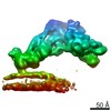
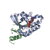
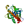
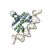
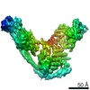
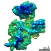
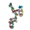
| Title | human complex II-BATS bound to membrane-attached Rab5a-GTP | ||||||||||||||||||||||||
 Components Components |
| ||||||||||||||||||||||||
 Keywords Keywords | ENDOCYTOSIS / Rab GTPase / Phosphatidylinositol 3-kinase | ||||||||||||||||||||||||
| Function / homology |  Function and homology information Function and homology informationregulation of protein serine/threonine kinase activity / lytic vacuole / maintenance of Golgi location / regulation of endosome size / nucleus-vacuole junction / cytoplasmic side of early endosome membrane / cellular response to aluminum ion / positive regulation of protein lipidation / protein localization to early endosome / postsynaptic endosome ...regulation of protein serine/threonine kinase activity / lytic vacuole / maintenance of Golgi location / regulation of endosome size / nucleus-vacuole junction / cytoplasmic side of early endosome membrane / cellular response to aluminum ion / positive regulation of protein lipidation / protein localization to early endosome / postsynaptic endosome / Toll Like Receptor 9 (TLR9) Cascade / positive regulation of stress granule assembly / Synthesis of PIPs at the late endosome membrane / phosphatidylinositol 3-kinase complex, class III / cellular response to oxygen-glucose deprivation / Synthesis of PIPs at the early endosome membrane / phosphatidylinositol 3-kinase complex, class III, type II / phosphatidylinositol 3-kinase complex, class III, type I / SARS-CoV-2 modulates autophagy / amyloid-beta clearance by transcytosis / response to mitochondrial depolarisation / synaptic vesicle recycling / presynaptic endosome / positive regulation of attachment of mitotic spindle microtubules to kinetochore / host-mediated activation of viral genome replication / plasma membrane to endosome transport / engulfment of apoptotic cell / negative regulation of lysosome organization / host-mediated perturbation of viral process / phosphatidylinositol kinase activity / positive regulation of autophagosome assembly / regulation of filopodium assembly / multivesicular body sorting pathway / Synthesis of PIPs at the Golgi membrane / cytoplasmic side of mitochondrial outer membrane / early endosome to late endosome transport / protein localization to phagophore assembly site / negative regulation of autophagosome assembly / receptor catabolic process / RAB geranylgeranylation / response to L-leucine / SMAD protein signal transduction / protein targeting to vacuole / late endosome to vacuole transport / protein targeting to lysosome / regulation of autophagosome assembly / endosome organization / pexophagy / RAB GEFs exchange GTP for GDP on RABs / early phagosome / retrograde vesicle-mediated transport, Golgi to endoplasmic reticulum / double-strand break repair via classical nonhomologous end joining / positive regulation of natural killer cell mediated cytotoxicity / phagophore assembly site / Translation of Replicase and Assembly of the Replication Transcription Complex / positive regulation of autophagosome maturation / TBC/RABGAPs / cellular response to nitrogen starvation / spindle organization / centrosome cycle / phosphatidylinositol 3-kinase / phosphatidylinositol-3-phosphate biosynthetic process / endosomal transport / 1-phosphatidylinositol-3-kinase activity / negative regulation of programmed cell death / response to vitamin E / mitotic metaphase chromosome alignment / lysosome organization / Macroautophagy / response to iron(II) ion / positive regulation of cardiac muscle hypertrophy / SNARE complex assembly / regulation of synaptic vesicle exocytosis / RSV-host interactions / p38MAPK cascade / cytoplasmic pattern recognition receptor signaling pathway / phosphatidylinositol phosphate biosynthetic process / phosphatidylinositol-mediated signaling / Synthesis of PIPs at the plasma membrane / autolysosome / positive regulation of exocytosis / Respiratory syncytial virus (RSV) attachment and entry / autophagosome membrane / PI3K Cascade / amyloid-beta metabolic process / protein secretion / chromosome, centromeric region / RHO GTPases Activate NADPH Oxidases / regulation of macroautophagy / canonical Wnt signaling pathway / autophagosome maturation / endocytic vesicle / axoneme / neuron development / synaptic vesicle endocytosis / cellular defense response / phagocytosis / autophagosome assembly / intercellular bridge / phosphatidylinositol 3-kinase binding Similarity search - Function | ||||||||||||||||||||||||
| Biological species |  Homo sapiens (human) Homo sapiens (human) | ||||||||||||||||||||||||
| Method | ELECTRON MICROSCOPY / subtomogram averaging / cryo EM / Resolution: 9.8 Å | ||||||||||||||||||||||||
 Authors Authors | Tremel, S. / Morado, D.R. / Kovtun, O. / Williams, R.L. / Briggs, J.A.G. / Munro, S. / Ohashi, Y. / Bertram, J. / Perisic, O. | ||||||||||||||||||||||||
| Funding support |  United Kingdom, 7items United Kingdom, 7items
| ||||||||||||||||||||||||
 Citation Citation | Journal: Nat Commun / Year: 2021 Title: Structural basis for VPS34 kinase activation by Rab1 and Rab5 on membranes. Authors: Shirley Tremel / Yohei Ohashi / Dustin R Morado / Jessie Bertram / Olga Perisic / Laura T L Brandt / Marie-Kristin von Wrisberg / Zhuo A Chen / Sarah L Maslen / Oleksiy Kovtun / Mark Skehel ...Authors: Shirley Tremel / Yohei Ohashi / Dustin R Morado / Jessie Bertram / Olga Perisic / Laura T L Brandt / Marie-Kristin von Wrisberg / Zhuo A Chen / Sarah L Maslen / Oleksiy Kovtun / Mark Skehel / Juri Rappsilber / Kathrin Lang / Sean Munro / John A G Briggs / Roger L Williams /   Abstract: The lipid phosphatidylinositol-3-phosphate (PI3P) is a regulator of two fundamental but distinct cellular processes, endocytosis and autophagy, so its generation needs to be under precise temporal ...The lipid phosphatidylinositol-3-phosphate (PI3P) is a regulator of two fundamental but distinct cellular processes, endocytosis and autophagy, so its generation needs to be under precise temporal and spatial control. PI3P is generated by two complexes that both contain the lipid kinase VPS34: complex II on endosomes (VPS34/VPS15/Beclin 1/UVRAG), and complex I on autophagosomes (VPS34/VPS15/Beclin 1/ATG14L). The endosomal GTPase Rab5 binds complex II, but the mechanism of VPS34 activation by Rab5 has remained elusive, and no GTPase is known to bind complex I. Here we show that Rab5a-GTP recruits endocytic complex II to membranes and activates it by binding between the VPS34 C2 and VPS15 WD40 domains. Electron cryotomography of complex II on Rab5a-decorated vesicles shows that the VPS34 kinase domain is released from inhibition by VPS15 and hovers over the lipid bilayer, poised for catalysis. We also show that the GTPase Rab1a, which is known to be involved in autophagy, recruits and activates the autophagy-specific complex I, but not complex II. Both Rabs bind to the same VPS34 interface but in a manner unique for each. These findings reveal how VPS34 complexes are activated on membranes by specific Rab GTPases and how they are recruited to unique cellular locations. | ||||||||||||||||||||||||
| History |
|
- Structure visualization
| Movie |
Movie viewer |
|---|---|
| Structure viewer | Molecule: MolmilJmol/JSmol |
- Downloads & links
-Download
| PDBx/mmCIF format | 7bl1.cif.gz | 501.3 KB | Display | PDBx/mmCIF format |
|---|---|---|---|---|
| PDB format | pdb7bl1.ent.gz | Display | PDB format | |
| PDBx/mmJSON format | 7bl1.json.gz | Tree view | PDBx/mmJSON format | |
| Others |  Other downloads Other downloads |
-Validation report
| Arichive directory | https://data.pdbj.org/pub/pdb/validation_reports/bl/7bl1ftp://data.pdbj.org/pub/pdb/validation_reports/bl/7bl1 | HTTPS FTP |
|---|
-Related structure data
| Related structure data | 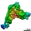 12214MC M: map data used to model this data C: citing same article ( |
|---|---|
| Similar structure data |


-Links
 PDBj
PDBj


- Assembly
| Deposited unit | 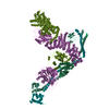
|
|---|---|
| 1 |
|
-Components
-Protein , 5 types, 5 molecules AAABBBCCCEEEDDD
| #1: Protein | Mass: 78258.836 Da / Num. of mol.: 1 Source method: isolated from a genetically manipulated source Source: (gene. exp.) Homo sapiens (human) / Gene: UVRAG / Cell line (production host): HEK293T / Production host: Homo sapiens (human) / References: UniProt: Q9P2Y5 |
|---|---|
| #2: Protein | Mass: 101680.328 Da / Num. of mol.: 1 Source method: isolated from a genetically manipulated source Source: (gene. exp.) Homo sapiens (human) / Gene: PIK3C3, VPS34 / Cell line (production host): expi293F / Production host: Homo sapiens (human) / References: UniProt: Q8NEB9, phosphatidylinositol 3-kinase |
| #3: Protein | Mass: 154790.391 Da / Num. of mol.: 1 Source method: isolated from a genetically manipulated source Source: (gene. exp.) Homo sapiens (human) / Gene: PIK3R4, VPS15 / Cell line (production host): expi293F / Production host: Homo sapiens (human)References: UniProt: Q99570, non-specific serine/threonine protein kinase |
| #4: Protein | Mass: 51953.102 Da / Num. of mol.: 1 Source method: isolated from a genetically manipulated source Source: (gene. exp.) Homo sapiens (human) / Gene: BECN1, GT197 / Cell line (production host): expi293F / Production host: Homo sapiens (human) / References: UniProt: Q14457 |
| #6: Protein | Mass: 18769.314 Da / Num. of mol.: 1 Source method: isolated from a genetically manipulated source Source: (gene. exp.) Homo sapiens (human) / Gene: RAB5A, RAB5Production host:  References: UniProt: P20339, small monomeric GTPase |
-Protein/peptide , 1 types, 1 molecules FFF
| #5: Protein/peptide | Mass: 1581.727 Da / Num. of mol.: 1 Source method: isolated from a genetically manipulated source Source: (gene. exp.) Homo sapiens (human) / Cell line (production host): expi293F / Production host: Homo sapiens (human) |
|---|
-Non-polymers , 2 types, 2 molecules 

| #7: Chemical | ChemComp-GTP /  Mass: 523.180 Da / Num. of mol.: 1 / Source method: obtained synthetically / Formula: C10H16N5O14P3 / Comment: GTP, energy-carrying molecule*YM Mass: 523.180 Da / Num. of mol.: 1 / Source method: obtained synthetically / Formula: C10H16N5O14P3 / Comment: GTP, energy-carrying molecule*YM |
|---|---|
| #8: Chemical | ChemComp-MG /  Mass: 24.305 Da / Num. of mol.: 1 / Source method: obtained synthetically / Formula: Mg Mass: 24.305 Da / Num. of mol.: 1 / Source method: obtained synthetically / Formula: Mg |
-Details
| Has ligand of interest | N |
|---|
-Experimental details
-Experiment
| Experiment | Method: ELECTRON MICROSCOPY |
|---|---|
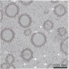
| EM experiment | Aggregation state: 3D ARRAY / 3D reconstruction method: subtomogram averaging |
- Sample preparation
| Component |
| ||||||||||||||||||||||||||||
|---|---|---|---|---|---|---|---|---|---|---|---|---|---|---|---|---|---|---|---|---|---|---|---|---|---|---|---|---|---|
| Molecular weight | Value: 392615 kDa/nm / Experimental value: NO | ||||||||||||||||||||||||||||
| Source (natural) |
| ||||||||||||||||||||||||||||
| Source (recombinant) |
| ||||||||||||||||||||||||||||
| Buffer solution | pH: 8 | ||||||||||||||||||||||||||||
| Buffer component |
| ||||||||||||||||||||||||||||
| Specimen | Conc.: 6.2 mg/ml / Embedding applied: NO / Shadowing applied: NO / Staining applied: NO / Vitrification applied: YES | ||||||||||||||||||||||||||||
| Specimen support | Details: Quorum SC7620 / Grid material: GOLD / Grid mesh size: 300 divisions/in. / Grid type: Quantifoil | ||||||||||||||||||||||||||||
| Vitrification | Instrument: FEI VITROBOT MARK I / Cryogen name: ETHANE / Humidity: 100 % / Chamber temperature: 316 K / Details: Blot force 20, blot time 6 s |
- Electron microscopy imaging
| Experimental equipment |  Model: Titan Krios / Image courtesy: FEI Company |
|---|---|
| Microscopy | Model: FEI TITAN KRIOS |
| Electron gun | Electron source:  FIELD EMISSION GUN / Accelerating voltage: 300 kV / Illumination mode: FLOOD BEAM FIELD EMISSION GUN / Accelerating voltage: 300 kV / Illumination mode: FLOOD BEAM |
| Electron lens | Mode: BRIGHT FIELD / Nominal magnification: 81000 X / Nominal defocus max: 5000 nm / Nominal defocus min: 2000 nm / Cs: 2.7 mm |
| Specimen holder | Cryogen: NITROGEN / Specimen holder model: FEI TITAN KRIOS AUTOGRID HOLDER |
| Image recording | Average exposure time: 0.55 sec. / Electron dose: 2.99 e/Å2 / Film or detector model: GATAN K3 BIOQUANTUM (6k x 4k) / Num. of grids imaged: 1 |
| Image scans | Width: 6000 / Height: 4000 |
- Processing
| Software | Name: REFMAC / Version: 5.8.0272 / Classification: refinement | |||||||||||||||||||||||||||||||||||||||||||||||||||||||||||||||||||||||||||||||||||||||||||||||||||||||||||||||||||||||||||||||||||||||||||||||||||
|---|---|---|---|---|---|---|---|---|---|---|---|---|---|---|---|---|---|---|---|---|---|---|---|---|---|---|---|---|---|---|---|---|---|---|---|---|---|---|---|---|---|---|---|---|---|---|---|---|---|---|---|---|---|---|---|---|---|---|---|---|---|---|---|---|---|---|---|---|---|---|---|---|---|---|---|---|---|---|---|---|---|---|---|---|---|---|---|---|---|---|---|---|---|---|---|---|---|---|---|---|---|---|---|---|---|---|---|---|---|---|---|---|---|---|---|---|---|---|---|---|---|---|---|---|---|---|---|---|---|---|---|---|---|---|---|---|---|---|---|---|---|---|---|---|---|---|---|---|
| EM software |
| |||||||||||||||||||||||||||||||||||||||||||||||||||||||||||||||||||||||||||||||||||||||||||||||||||||||||||||||||||||||||||||||||||||||||||||||||||
| CTF correction | Type: PHASE FLIPPING AND AMPLITUDE CORRECTION | |||||||||||||||||||||||||||||||||||||||||||||||||||||||||||||||||||||||||||||||||||||||||||||||||||||||||||||||||||||||||||||||||||||||||||||||||||
| Symmetry | Point symmetry: C1 (asymmetric) | |||||||||||||||||||||||||||||||||||||||||||||||||||||||||||||||||||||||||||||||||||||||||||||||||||||||||||||||||||||||||||||||||||||||||||||||||||
| 3D reconstruction | Resolution: 9.8 Å / Resolution method: FSC 0.143 CUT-OFF / Num. of particles: 26979 / Algorithm: FOURIER SPACE / Symmetry type: POINT | |||||||||||||||||||||||||||||||||||||||||||||||||||||||||||||||||||||||||||||||||||||||||||||||||||||||||||||||||||||||||||||||||||||||||||||||||||
| EM volume selection | Num. of tomograms: 105 / Num. of volumes extracted: 191196 / Reference model: two Gaussian filtered elipsoids | |||||||||||||||||||||||||||||||||||||||||||||||||||||||||||||||||||||||||||||||||||||||||||||||||||||||||||||||||||||||||||||||||||||||||||||||||||
| Atomic model building | B value: 320 / Protocol: FLEXIBLE FIT / Space: REAL / Target criteria: correlation Details: An initial model was build with SWISSMODEL. using 5DFZ, 4DDP and 3IHY. This was then fit manually to density while running restrained MD with ISOLDE. The model was refined in REFMAC with ...Details: An initial model was build with SWISSMODEL. using 5DFZ, 4DDP and 3IHY. This was then fit manually to density while running restrained MD with ISOLDE. The model was refined in REFMAC with self-restraints (4.3) and restraints to 3MJH, 4DDP and 3IHY. | |||||||||||||||||||||||||||||||||||||||||||||||||||||||||||||||||||||||||||||||||||||||||||||||||||||||||||||||||||||||||||||||||||||||||||||||||||
| Atomic model building | 3D fitting-ID: 1 / Source name: PDB / Type: experimental model
| |||||||||||||||||||||||||||||||||||||||||||||||||||||||||||||||||||||||||||||||||||||||||||||||||||||||||||||||||||||||||||||||||||||||||||||||||||
| Refinement | Resolution: 9.8→234.63 Å / Cor.coef. Fo:Fc: 0.954 / WRfactor Rwork: 0.1573 / SU B: 303.369 / SU ML: 1.88 / Average fsc overall: 0.9814 / Average fsc work: 0.9814 Details: Hydrogens have been used if present in the input file
| |||||||||||||||||||||||||||||||||||||||||||||||||||||||||||||||||||||||||||||||||||||||||||||||||||||||||||||||||||||||||||||||||||||||||||||||||||
| Solvent computation | Solvent model: BABINET MODEL | |||||||||||||||||||||||||||||||||||||||||||||||||||||||||||||||||||||||||||||||||||||||||||||||||||||||||||||||||||||||||||||||||||||||||||||||||||
| Displacement parameters | Biso mean: 320.935 Å2
| |||||||||||||||||||||||||||||||||||||||||||||||||||||||||||||||||||||||||||||||||||||||||||||||||||||||||||||||||||||||||||||||||||||||||||||||||||
| Refine LS restraints |
| |||||||||||||||||||||||||||||||||||||||||||||||||||||||||||||||||||||||||||||||||||||||||||||||||||||||||||||||||||||||||||||||||||||||||||||||||||
| LS refinement shell | Refine-ID: ELECTRON MICROSCOPY / Num. reflection Rfree: _ / Total num. of bins used: 20 / % reflection obs: 100 %
|

