[English] 日本語
Yorodumi
- PDB-3ihy: Human PIK3C3 crystal structure -

+
Open data


ID or keywords:

Loading...

-
Basic information

Entry
Database: PDB / ID: 3ihy
TitleHuman PIK3C3 crystal structure
ComponentsPhosphatidylinositol 3-kinase catalytic subunit type 3
KeywordsTRANSFERASE / Alpha/Beta protein / Structural Genomics / SGC Stockholm / Structural Genomics Consortium / SGC / ATP-binding / Kinase / Manganese / Nucleotide-binding / Phosphoprotein
Function / homology
Function and homology information


Toll Like Receptor 9 (TLR9) Cascade / protein lipidation / Synthesis of PIPs at the late endosome membrane / Synthesis of PIPs at the early endosome membrane / phosphatidylinositol 3-kinase complex, class III / phosphatidylinositol 3-kinase complex, class III, type II / phosphatidylinositol 3-kinase complex, class III, type I / positive regulation by host of viral genome replication / Synthesis of PIPs at the Golgi membrane / phosphatidylinositol kinase activity ...Toll Like Receptor 9 (TLR9) Cascade / protein lipidation / Synthesis of PIPs at the late endosome membrane / Synthesis of PIPs at the early endosome membrane / phosphatidylinositol 3-kinase complex, class III / phosphatidylinositol 3-kinase complex, class III, type II / phosphatidylinositol 3-kinase complex, class III, type I / positive regulation by host of viral genome replication / Synthesis of PIPs at the Golgi membrane / phosphatidylinositol kinase activity / protein localization to phagophore assembly site / protein targeting to lysosome / early endosome to late endosome transport / phagophore assembly site / phosphatidylinositol-mediated signaling / Translation of Replicase and Assembly of the Replication Transcription Complex / autolysosome / phosphatidylinositol 3-kinase / phosphatidylinositol-3-phosphate biosynthetic process / 1-phosphatidylinositol-3-kinase activity / Macroautophagy / axoneme / autophagosome maturation / phosphatidylinositol phosphate biosynthetic process / autophagosome assembly / RHO GTPases Activate NADPH Oxidases / PI3K Cascade / regulation of macroautophagy / cellular response to glucose starvation / regulation of cytokinesis / regulation of autophagy / Antigen Presentation: Folding, assembly and peptide loading of class I MHC / macroautophagy / autophagy / peroxisome / endocytosis / phagocytic vesicle membrane / late endosome / Translation of Replicase and Assembly of the Replication Transcription Complex / kinase activity / midbody / endosome / protein kinase activity / cell cycle / cell division / phosphorylation / SARS-CoV-2 activates/modulates innate and adaptive immune responses / ATP binding / membrane / cytosol / cytoplasm
Similarity search - Function
Phosphatidylinositol 3-kinase, Vps34 type / Phosphatidylinositol 3-/4-kinase, catalytic domain / Phosphatidylinositol 3-kinase, accessory domain (PIK) / Phosphatidylinositol 3-kinase Catalytic Subunit; Chain A, domain 4 / Phosphatidylinositol 3-kinase Catalytic Subunit; Chain A, domain 4 / Phosphatidylinositol 3-kinase Catalytic Subunit; Chain A, Domain 5 / Phosphoinositide 3-kinase C2 / Phosphoinositide 3-kinase, region postulated to contain C2 domain / C2 phosphatidylinositol 3-kinase-type domain / C2 phosphatidylinositol 3-kinase (PI3K)-type domain profile. ...Phosphatidylinositol 3-kinase, Vps34 type / Phosphatidylinositol 3-/4-kinase, catalytic domain / Phosphatidylinositol 3-kinase, accessory domain (PIK) / Phosphatidylinositol 3-kinase Catalytic Subunit; Chain A, domain 4 / Phosphatidylinositol 3-kinase Catalytic Subunit; Chain A, domain 4 / Phosphatidylinositol 3-kinase Catalytic Subunit; Chain A, Domain 5 / Phosphoinositide 3-kinase C2 / Phosphoinositide 3-kinase, region postulated to contain C2 domain / C2 phosphatidylinositol 3-kinase-type domain / C2 phosphatidylinositol 3-kinase (PI3K)-type domain profile. / Phosphoinositide 3-kinase, accessory (PIK) domain superfamily / Phosphoinositide 3-kinase family, accessory domain (PIK domain) / Phosphoinositide 3-kinase family, accessory domain (PIK domain) / Phosphoinositide 3-kinase, accessory (PIK) domain / Phosphatidylinositol kinase / PIK helical domain profile. / Phosphatidylinositol 3- and 4-kinases signature 1. / Phosphatidylinositol 3/4-kinase, conserved site / Phosphatidylinositol 3- and 4-kinases signature 2. / Phosphatidylinositol 3-/4-kinase, catalytic domain superfamily / Phosphoinositide 3-kinase, catalytic domain / Phosphatidylinositol 3- and 4-kinase / Phosphatidylinositol 3- and 4-kinases catalytic domain profile. / Phosphatidylinositol 3-/4-kinase, catalytic domain / C2 domain superfamily / Serine Threonine Protein Phosphatase 5, Tetratricopeptide repeat / Alpha Horseshoe / Armadillo-type fold / Protein kinase-like domain superfamily / 2-Layer Sandwich / Orthogonal Bundle / Mainly Alpha / Alpha Beta
Similarity search - Domain/homology
Phosphatidylinositol 3-kinase catalytic subunit type 3
Similarity search - Component
Biological speciesHomo sapiens (human)
MethodX-RAY DIFFRACTION / SYNCHROTRON / MOLECULAR REPLACEMENT / Resolution: 2.8 Å
AuthorsSiponen, M.I. / Tresaugues, L. / Arrowsmith, C.H. / Berglund, H. / Bountra, C. / Collins, R. / Edwards, A.M. / Flodin, S. / Flores, A. / Graslund, S. ...Siponen, M.I. / Tresaugues, L. / Arrowsmith, C.H. / Berglund, H. / Bountra, C. / Collins, R. / Edwards, A.M. / Flodin, S. / Flores, A. / Graslund, S. / Hammarstrom, M. / Johansson, A. / Johansson, I. / Karlberg, T. / Kotenyova, T. / Kotzsch, A. / Kragh Nielsen, T. / Moche, M. / Nyman, T. / Persson, C. / Roos, A.K. / Sagemark, J. / Schueler, H. / Schutz, P. / Thorsell, A.G. / Van Den Berg, S. / Weigelt, J. / Welin, M. / Wisniewska, M. / Nordlund, P. / Structural Genomics Consortium (SGC)
CitationJournal: TO BE PUBLISHED
Title: Human PIK3C3 crystal structure
Authors: Siponen, M.I. / Tresaugues, L. / Arrowsmith, C.H. / Berglund, H. / Bountra, C. / Collins, R. / Edwards, A.M. / Flodin, S. / Flores, A. / Graslund, S. / Hammarstrom, M. / Johansson, A. / ...Authors: Siponen, M.I. / Tresaugues, L. / Arrowsmith, C.H. / Berglund, H. / Bountra, C. / Collins, R. / Edwards, A.M. / Flodin, S. / Flores, A. / Graslund, S. / Hammarstrom, M. / Johansson, A. / Johansson, I. / Karlberg, T. / Kotenyova, T. / Kotzsch, A. / Kragh Nielsen, T. / Moche, M. / Nyman, T. / Persson, C. / Roos, A.K. / Sagemark, J. / Schueler, H. / Schutz, P. / Thorsell, A.G. / Van Den Berg, S. / Weigelt, J. / Welin, M. / Wisniewska, M. / Nordlund, P.
History
DepositionJul 31, 2009Deposition site: RCSB / Processing site: RCSB
Revision 1.0Oct 13, 2009Provider: repository / Type: Initial release
Revision 1.1Jul 13, 2011Group: Version format compliance
Revision 1.2Jan 24, 2018Group: Database references / Category: citation_author / Item: _citation_author.name
Revision 1.3Feb 21, 2024Group: Data collection / Database references
Category: chem_comp_atom / chem_comp_bond ...chem_comp_atom / chem_comp_bond / database_2 / struct_ref_seq_dif
Item: _database_2.pdbx_DOI / _database_2.pdbx_database_accession / _struct_ref_seq_dif.details

-
Structure visualization

Structure viewerMolecule:
MolmilJmol/JSmol

Downloads & links

-
Assembly

Deposited unit
A: Phosphatidylinositol 3-kinase catalytic subunit type 3
B: Phosphatidylinositol 3-kinase catalytic subunit type 3
C: Phosphatidylinositol 3-kinase catalytic subunit type 3
D: Phosphatidylinositol 3-kinase catalytic subunit type 3
E: Phosphatidylinositol 3-kinase catalytic subunit type 3


Theoretical massNumber of molelcules
Total (without water)342,7825
Polymers342,7825
Non-polymers00
Water181
1
A: Phosphatidylinositol 3-kinase catalytic subunit type 3


Theoretical massNumber of molelcules
Total (without water)68,5561
Polymers68,5561
Non-polymers00
Water0
TypeNameSymmetry operationNumber
identity operation1_555x,y,z1
MethodPISA
2
B: Phosphatidylinositol 3-kinase catalytic subunit type 3


Theoretical massNumber of molelcules
Total (without water)68,5561
Polymers68,5561
Non-polymers00
Water181
TypeNameSymmetry operationNumber
identity operation1_555x,y,z1
MethodPISA
3
C: Phosphatidylinositol 3-kinase catalytic subunit type 3


Theoretical massNumber of molelcules
Total (without water)68,5561
Polymers68,5561
Non-polymers00
Water0
TypeNameSymmetry operationNumber
identity operation1_555x,y,z1
MethodPISA
4
D: Phosphatidylinositol 3-kinase catalytic subunit type 3


Theoretical massNumber of molelcules
Total (without water)68,5561
Polymers68,5561
Non-polymers00
Water0
TypeNameSymmetry operationNumber
identity operation1_555x,y,z1
MethodPISA
5
E: Phosphatidylinositol 3-kinase catalytic subunit type 3


Theoretical massNumber of molelcules
Total (without water)68,5561
Polymers68,5561
Non-polymers00
Water0
TypeNameSymmetry operationNumber
identity operation1_555x,y,z1
MethodPISA
Unit cell
Length a, b, c (Å)62.297, 96.491, 150.679
Angle α, β, γ (deg.)107.77, 91.75, 92.41
Int Tables number1
Space group name H-MP1

-
Components

#1: Protein
Phosphatidylinositol 3-kinase catalytic subunit type 3 / PtdIns-3-kinase type 3 / PI3-kinase type 3 / PI3K type 3 / Phosphoinositide-3-kinase class 3 / ...PtdIns-3-kinase type 3 / PI3-kinase type 3 / PI3K type 3 / Phosphoinositide-3-kinase class 3 / Phosphatidylinositol 3-kinase p100 subunit


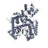
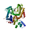
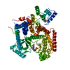
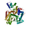
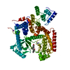
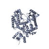
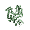
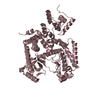
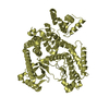
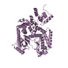
Mass: 68556.336 Da / Num. of mol.: 5 / Fragment: UNP residues 282-879
Source method: isolated from a genetically manipulated source
Source: (gene. exp.) Homo sapiens (human) / Gene: PIK3C3, PIK3C3A / Plasmid: pNIC Bsa4 / Production host: Escherichia coli (E. coli) / Strain (production host): BL21(DE3) R3 pRARE / References: UniProt: Q8NEB9, phosphatidylinositol 3-kinase
#2: Water ChemComp-HOH / water / Water


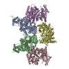
Mass: 18.015 Da / Num. of mol.: 1 / Source method: isolated from a natural source / Formula: H2O

-
Experimental details

-
Experiment

ExperimentMethod: X-RAY DIFFRACTION / Number of used crystals: 1

-
Sample preparation

CrystalDensity Matthews: 2.51 Å3/Da / Density % sol: 51.02 %
Crystal growTemperature: 277 K / Method: vapor diffusion, sitting drop / pH: 8.5
Details: 0.2M ammonium acetate 25% PEG3350 0.1M Tris, pH 8.5, VAPOR DIFFUSION, SITTING DROP, temperature 277K

-
Data collection

DiffractionMean temperature: 100 K
Diffraction sourceSource: SYNCHROTRON / Site: ESRF / Beamline: ID14-2
DetectorType: ADSC QUANTUM 4 / Detector: CCD / Date: Apr 4, 2009 / Details: mirrors
RadiationMonochromator: si 111 / Protocol: SINGLE WAVELENGTH / Monochromatic (M) / Laue (L): M / Scattering type: x-ray
Radiation wavelengthRelative weight: 1
ReflectionResolution: 2.8→19.84 Å

-
Processing

Software
NameVersionClassification
ADSCQuantumdata collection
PHASERphasing
REFMAC5.2.0019refinement
XDSdata reduction
SCALAdata scaling
RefinementMethod to determine structure: MOLECULAR REPLACEMENT / Resolution: 2.8→19.84 Å / Cor.coef. Fo:Fc: 0.938 / Cor.coef. Fo:Fc free: 0.88 / SU B: 14.662 / SU ML: 0.285 / Cross valid method: THROUGHOUT / σ(F): 0 / ESU R Free: 0.387 / Stereochemistry target values: MAXIMUM LIKELIHOOD / Details: HYDROGENS HAVE BEEN ADDED IN THE RIDING POSITIONS
RfactorNum. reflection% reflectionSelection details
Rfree0.25505 4076 5 %RANDOM
Rwork0.18663 ---
obs0.19006 77091 99.04 %-
Solvent computationIon probe radii: 0.8 Å / Shrinkage radii: 0.8 Å / VDW probe radii: 1.2 Å / Solvent model: MASK
Displacement parametersBiso mean: 33.483 Å2
Baniso -1Baniso -2Baniso -3
1-0 Å20.01 Å20.01 Å2
2---0.01 Å2-0.01 Å2
3---0 Å2
Refine analyzeLuzzati coordinate error free: 0.285 Å
Refinement stepCycle: LAST / Resolution: 2.8→19.84 Å
ProteinNucleic acidLigandSolventTotal
Num. atoms21669 0 0 1 21670
Refine LS restraints
Refine-IDTypeDev idealDev ideal targetNumber
X-RAY DIFFRACTIONr_bond_refined_d0.0140.02222121
X-RAY DIFFRACTIONr_bond_other_d0.0060.0215313
X-RAY DIFFRACTIONr_angle_refined_deg1.5031.98229894
X-RAY DIFFRACTIONr_angle_other_deg0.96337483
X-RAY DIFFRACTIONr_dihedral_angle_1_deg6.26552665
X-RAY DIFFRACTIONr_dihedral_angle_2_deg39.524.7541056
X-RAY DIFFRACTIONr_dihedral_angle_3_deg19.63154153
X-RAY DIFFRACTIONr_dihedral_angle_4_deg19.47815130
X-RAY DIFFRACTIONr_chiral_restr0.0780.23344
X-RAY DIFFRACTIONr_gen_planes_refined0.0040.0224136
X-RAY DIFFRACTIONr_gen_planes_other0.0010.024254
X-RAY DIFFRACTIONr_nbd_refined0.2360.25444
X-RAY DIFFRACTIONr_nbd_other0.2040.215933
X-RAY DIFFRACTIONr_nbtor_refined0.190.210898
X-RAY DIFFRACTIONr_nbtor_other0.0940.211901
X-RAY DIFFRACTIONr_xyhbond_nbd_refined0.1710.2445
X-RAY DIFFRACTIONr_xyhbond_nbd_other0.0640.23
X-RAY DIFFRACTIONr_metal_ion_refined
X-RAY DIFFRACTIONr_metal_ion_other
X-RAY DIFFRACTIONr_symmetry_vdw_refined0.240.257
X-RAY DIFFRACTIONr_symmetry_vdw_other0.2770.2128
X-RAY DIFFRACTIONr_symmetry_hbond_refined0.1730.211
X-RAY DIFFRACTIONr_symmetry_hbond_other
X-RAY DIFFRACTIONr_symmetry_metal_ion_refined
X-RAY DIFFRACTIONr_symmetry_metal_ion_other
X-RAY DIFFRACTIONr_mcbond_it1.1081.517162
X-RAY DIFFRACTIONr_mcbond_other0.0831.55336
X-RAY DIFFRACTIONr_mcangle_it1.018221670
X-RAY DIFFRACTIONr_scbond_it1.818310048
X-RAY DIFFRACTIONr_scangle_it2.614.58224
X-RAY DIFFRACTIONr_rigid_bond_restr
X-RAY DIFFRACTIONr_sphericity_free
X-RAY DIFFRACTIONr_sphericity_bonded
LS refinement shellResolution: 2.8→2.871 Å / Total num. of bins used: 20
RfactorNum. reflection% reflection
Rfree0.373 275 -
Rwork0.282 5633 -
obs--98.47 %

+
About Yorodumi

-
News

-
Feb 9, 2022. New format data for meta-information of EMDB entries

New format data for meta-information of EMDB entries

  • Version 3 of the EMDB header file is now the official format.
  • The previous official version 1.9 will be removed from the archive.

Related info.:EMDB header

External links:wwPDB to switch to version 3 of the EMDB data model

-
Aug 12, 2020. Covid-19 info

Covid-19 info

URL: https://pdbj.org/emnavi/covid19.php

New page: Covid-19 featured information page in EM Navigator.

Related info.:Covid-19 info / Mar 5, 2020. Novel coronavirus structure data

+
Mar 5, 2020. Novel coronavirus structure data

Novel coronavirus structure data

Related info.:Yorodumi Speices / Aug 12, 2020. Covid-19 info

External links:COVID-19 featured content - PDBj / Molecule of the Month (242):Coronavirus Proteases

+
Jan 31, 2019. EMDB accession codes are about to change! (news from PDBe EMDB page)

EMDB accession codes are about to change! (news from PDBe EMDB page)

  • The allocation of 4 digits for EMDB accession codes will soon come to an end. Whilst these codes will remain in use, new EMDB accession codes will include an additional digit and will expand incrementally as the available range of codes is exhausted. The current 4-digit format prefixed with “EMD-” (i.e. EMD-XXXX) will advance to a 5-digit format (i.e. EMD-XXXXX), and so on. It is currently estimated that the 4-digit codes will be depleted around Spring 2019, at which point the 5-digit format will come into force.
  • The EM Navigator/Yorodumi systems omit the EMD- prefix.

Related info.:Q: What is EMD? / ID/Accession-code notation in Yorodumi/EM Navigator

External links:EMDB Accession Codes are Changing Soon! / Contact to PDBj

+
Jul 12, 2017. Major update of PDB

Major update of PDB

  • wwPDB released updated PDB data conforming to the new PDBx/mmCIF dictionary.
  • This is a major update changing the version number from 4 to 5, and with Remediation, in which all the entries are updated.
  • In this update, many items about electron microscopy experimental information are reorganized (e.g. em_software).
  • Now, EM Navigator and Yorodumi are based on the updated data.

External links:wwPDB Remediation / Enriched Model Files Conforming to OneDep Data Standards Now Available in the PDB FTP Archive

-
Yorodumi

Thousand views of thousand structures

  • Yorodumi is a browser for structure data from EMDB, PDB, SASBDB, etc.
  • This page is also the successor to EM Navigator detail page, and also detail information page/front-end page for Omokage search.
  • The word "yorodu" (or yorozu) is an old Japanese word meaning "ten thousand". "mi" (miru) is to see.

Related info.:EMDB / PDB / SASBDB / Comparison of 3 databanks / Yorodumi Search / Aug 31, 2016. New EM Navigator & Yorodumi / Yorodumi Papers / Jmol/JSmol / Function and homology information / Changes in new EM Navigator and Yorodumi

Read more