- PDB-4ph4: The crystal structure of Human VPS34 in complex with PIK-III -
+
Open data
ID or keywords:
Loading...
-
Basic information
Entry
Database: PDB / ID: 4ph4
Title
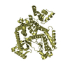
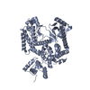
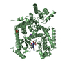
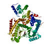
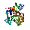
The crystal structure of Human VPS34 in complex with PIK-III
Components
Phosphatidylinositol 3-kinase catalytic subunit type 3
Keywords
TRANSFERASE/TRANSFERASE INHIBITOR / VPS34 / autophagy / class III / phosphatidylinositol-3-kinase / PIK3C3 / TRANSFERASE-TRANSFERASE INHIBITOR complex
Function / homology
Function and homology information
positive regulation of protein lipidation / postsynaptic endosome / Toll Like Receptor 9 (TLR9) Cascade / Synthesis of PIPs at the late endosome membrane / phosphatidylinositol 3-kinase complex, class III / Synthesis of PIPs at the early endosome membrane / phosphatidylinositol 3-kinase complex, class III, type II / phosphatidylinositol 3-kinase complex, class III, type I / presynaptic endosome / host-mediated activation of viral genome replication ...positive regulation of protein lipidation / postsynaptic endosome / Toll Like Receptor 9 (TLR9) Cascade / Synthesis of PIPs at the late endosome membrane / phosphatidylinositol 3-kinase complex, class III / Synthesis of PIPs at the early endosome membrane / phosphatidylinositol 3-kinase complex, class III, type II / phosphatidylinositol 3-kinase complex, class III, type I / presynaptic endosome / host-mediated activation of viral genome replication / phosphatidylinositol kinase activity / Synthesis of PIPs at the Golgi membrane / early endosome to late endosome transport / protein localization to phagophore assembly site / response to L-leucine / protein targeting to lysosome / endosome organization / pexophagy / positive regulation of natural killer cell mediated cytotoxicity / phagophore assembly site / Translation of Replicase and Assembly of the Replication Transcription Complex / phosphatidylinositol 3-kinase / phosphatidylinositol-3-phosphate biosynthetic process / 1-phosphatidylinositol-3-kinase activity / Macroautophagy / phosphatidylinositol phosphate biosynthetic process / phosphatidylinositol-mediated signaling / autolysosome / PI3K Cascade / regulation of macroautophagy / RHO GTPases Activate NADPH Oxidases / autophagosome maturation / axoneme / synaptic vesicle endocytosis / autophagosome assembly / cellular response to glucose starvation / autophagosome / regulation of autophagy / regulation of cytokinesis / Antigen Presentation: Folding, assembly and peptide loading of class I MHC / macroautophagy / phosphatidylinositol 3-kinase/protein kinase B signal transduction / protein processing / autophagy / GABA-ergic synapse / phagocytic vesicle membrane / endocytosis / late endosome / peroxisome / Translation of Replicase and Assembly of the Replication Transcription Complex / midbody / protein kinase activity / endosome / cell division / SARS-CoV-2 activates/modulates innate and adaptive immune responses / glutamatergic synapse / ATP binding / membrane / plasma membrane / cytoplasm / cytosol Similarity search - Function
Mass: 18.015 Da / Num. of mol.: 91 / Source method: isolated from a natural source / Formula: H2O
-
Experimental details
-
Experiment
Experiment
Method: X-RAY DIFFRACTION / Number of used crystals: 1
-
Sample preparation
Crystal
Density Matthews: 3.6 Å3/Da / Density % sol: 65.84 %
Crystal grow
Temperature: 303.15 K / Method: vapor diffusion, hanging drop Details: VPS34 protein and PIK-III were mixed and incubated on ice for 1 hr (final PIK-III concentration was 1 mM). Prior to crystallization, the mixture was passed through a 0.2 um filter. The ...Details: VPS34 protein and PIK-III were mixed and incubated on ice for 1 hr (final PIK-III concentration was 1 mM). Prior to crystallization, the mixture was passed through a 0.2 um filter. The protein:ligand complex was crystallized using the hanging drop vapor diffusion method in Nextal plates: 6 uL of protein solution was mixed with 4 uL of precipitant, which consisted of 20% (w/v) PEG 3350, 100 mM bis-tris propane, and 200 mM Na-K-phosphate. The resulting drop was suspended over a reservoir of 0.3 mL of precipitant and sealed with a screw cap. The crystals grew at 30 degC in approximately 12-24 hr.
-
Data collection
Diffraction
Mean temperature: 100 K
Diffraction source
Source: SYNCHROTRON / Site: ALS / Beamline: 5.0.2 / Wavelength: 0.9774 Å
In the structure databanks used in Yorodumi, some data are registered as the other names, "COVID-19 virus" and "2019-nCoV". Here are the details of the virus and the list of structure data.
Jan 31, 2019. EMDB accession codes are about to change! (news from PDBe EMDB page)
EMDB accession codes are about to change! (news from PDBe EMDB page)
The allocation of 4 digits for EMDB accession codes will soon come to an end. Whilst these codes will remain in use, new EMDB accession codes will include an additional digit and will expand incrementally as the available range of codes is exhausted. The current 4-digit format prefixed with “EMD-” (i.e. EMD-XXXX) will advance to a 5-digit format (i.e. EMD-XXXXX), and so on. It is currently estimated that the 4-digit codes will be depleted around Spring 2019, at which point the 5-digit format will come into force.
The EM Navigator/Yorodumi systems omit the EMD- prefix.
Related info.:Q: What is EMD? / ID/Accession-code notation in Yorodumi/EM Navigator
Yorodumi is a browser for structure data from EMDB, PDB, SASBDB, etc.
This page is also the successor to EM Navigator detail page, and also detail information page/front-end page for Omokage search.
The word "yorodu" (or yorozu) is an old Japanese word meaning "ten thousand". "mi" (miru) is to see.
Related info.:EMDB / PDB / SASBDB / Comparison of 3 databanks / Yorodumi Search / Aug 31, 2016. New EM Navigator & Yorodumi / Yorodumi Papers / Jmol/JSmol / Function and homology information / Changes in new EM Navigator and Yorodumi
 Movie
Movie Controller


 Open data
 Basic information
 Components
 Keywords
 Function and homology information
 Homo sapiens (human)
 X-RAY DIFFRACTION /
  Authors
 Citation
 Structure visualization
 Downloads & links
 Other downloads
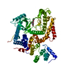


 PDBj
PDBj


 Assembly

 Spodoptera frugiperda (fall armyworm) / References: UniProt: Q8NEB9, phosphatidylinositol 3-kinase
 Mass: 92.094 Da / Num. of mol.: 1 / Source method: obtained synthetically / Formula: C3H8O3
 Mass: 319.364 Da / Num. of mol.: 1 / Source method: obtained synthetically / Formula: C17H17N7
 Mass: 18.015 Da / Num. of mol.: 91 / Source method: isolated from a natural source / Formula: H2O
 Sample preparation
 / Beamline: 5.0.2 / Wavelength: 0.9774 Å
 Processing