- PDB-7bii: Crystal structure of Nematocida HUWE1 -
+
Open data
ID or keywords:
Loading...
-
Basic information
Entry
Database: PDB / ID: 7bii
Title
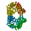
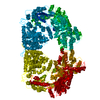
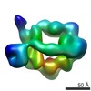
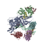
Crystal structure of Nematocida HUWE1
Components
E3 ubiquitin-protein ligase HUWE1
Keywords
LIGASE / HECT E3 ligase / ubiquitin / protein quality control
Function / homology
Function and homology information
HECT-type E3 ubiquitin transferase / ubiquitin protein ligase activity / ubiquitin-dependent protein catabolic process / protein ubiquitination / membrane / cytoplasm Similarity search - Function
E3 ubiquitin ligase, domain of unknown function DUF908 / E3 ubiquitin ligase, domain of unknown function DUF913 / Domain of Unknown Function (DUF908) / Domain of Unknown Function (DUF913) / : / HECT domain / HECT, E3 ligase catalytic domain / HECT-domain (ubiquitin-transferase) / HECT domain profile. / Domain Homologous to E6-AP Carboxyl Terminus with / Armadillo-type fold Similarity search - Domain/homology
In the structure databanks used in Yorodumi, some data are registered as the other names, "COVID-19 virus" and "2019-nCoV". Here are the details of the virus and the list of structure data.
Jan 31, 2019. EMDB accession codes are about to change! (news from PDBe EMDB page)
EMDB accession codes are about to change! (news from PDBe EMDB page)
The allocation of 4 digits for EMDB accession codes will soon come to an end. Whilst these codes will remain in use, new EMDB accession codes will include an additional digit and will expand incrementally as the available range of codes is exhausted. The current 4-digit format prefixed with “EMD-” (i.e. EMD-XXXX) will advance to a 5-digit format (i.e. EMD-XXXXX), and so on. It is currently estimated that the 4-digit codes will be depleted around Spring 2019, at which point the 5-digit format will come into force.
The EM Navigator/Yorodumi systems omit the EMD- prefix.
Related info.:Q: What is EMD? / ID/Accession-code notation in Yorodumi/EM Navigator
Yorodumi is a browser for structure data from EMDB, PDB, SASBDB, etc.
This page is also the successor to EM Navigator detail page, and also detail information page/front-end page for Omokage search.
The word "yorodu" (or yorozu) is an old Japanese word meaning "ten thousand". "mi" (miru) is to see.
Related info.:EMDB / PDB / SASBDB / Comparison of 3 databanks / Yorodumi Search / Aug 31, 2016. New EM Navigator & Yorodumi / Yorodumi Papers / Jmol/JSmol / Function and homology information / Changes in new EM Navigator and Yorodumi
 Movie
Movie Controller


 Open data
 Basic information
 Components
 Keywords
 Function and homology information
 Nematocida sp. ERTm5 (fungus)
 X-RAY DIFFRACTION /
  Authors
 Austria, 1items
  Citation
 Structure visualization
 Downloads & links
 Other downloads


 PDBj
PDBj
 Assembly
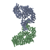


 Trichoplusia ni (cabbage looper) / References: UniProt: A0A177ELV2
 Sample preparation
 / Beamline: X06SA / Wavelength: 1 Å
 Processing