 Movie
Movie Controller


+ Open data

- Basic information
| Entry | Database: PDB / ID: 4q7j | ||||||
|---|---|---|---|---|---|---|---|
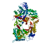
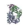
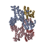
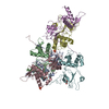
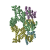
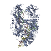
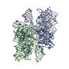
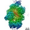
| Title | Complex structure of viral RNA polymerase | ||||||
 Components Components |
| ||||||
 Keywords Keywords | TRANSLATION/TRANSFERASE / RNA polymerase / RNA binding motif / RNA dependent RNA polymerization / TRANSLATION-TRANSFERASE complex | ||||||
| Function / homology |  Function and homology information Function and homology informationguanyl-nucleotide exchange factor complex / positive regulation of cytoplasmic translation / protein-synthesizing GTPase / guanosine tetraphosphate binding / translational elongation / translation elongation factor activity / negative regulation of cytoplasmic translation / guanyl-nucleotide exchange factor activity / ribosomal small subunit assembly / cytosolic small ribosomal subunit ...guanyl-nucleotide exchange factor complex / positive regulation of cytoplasmic translation / protein-synthesizing GTPase / guanosine tetraphosphate binding / translational elongation / translation elongation factor activity / negative regulation of cytoplasmic translation / guanyl-nucleotide exchange factor activity / ribosomal small subunit assembly / cytosolic small ribosomal subunit / cytoplasmic translation / single-stranded RNA binding / structural constituent of ribosome / translation / RNA-directed RNA polymerase / response to antibiotic / viral RNA genome replication / nucleotide binding / RNA-directed RNA polymerase activity / mRNA binding / GTPase activity / GTP binding / magnesium ion binding / RNA binding / zinc ion binding / membrane / metal ion binding / plasma membrane / cytosol / cytoplasm Similarity search - Function | ||||||
| Biological species |   Enterobacteria phage Qbeta (virus) Enterobacteria phage Qbeta (virus) | ||||||
| Method |  X-RAY DIFFRACTION / SYNCHROTRON / MOLECULAR REPLACEMENT / Resolution: 2.9 Å X-RAY DIFFRACTION / SYNCHROTRON / MOLECULAR REPLACEMENT / Resolution: 2.9 Å | ||||||
 Authors Authors | Takeshita, D. | ||||||
 Citation Citation | Journal: Nucleic Acids Res. / Year: 2014 Title: Molecular insights into replication initiation by Q beta replicase using ribosomal protein S1. Authors: Takeshita, D. / Yamashita, S. / Tomita, K. | ||||||
| History |
|
- Structure visualization
| Structure viewer | Molecule: MolmilJmol/JSmol |
|---|
- Downloads & links
-Download
| PDBx/mmCIF format | 4q7j.cif.gz | 535.4 KB | Display | PDBx/mmCIF format |
|---|---|---|---|---|
| PDB format | pdb4q7j.ent.gz | 434.5 KB | Display | PDB format |
| PDBx/mmJSON format | 4q7j.json.gz | Tree view | PDBx/mmJSON format | |
| Others |  Other downloads Other downloads |
-Validation report
| Arichive directory | https://data.pdbj.org/pub/pdb/validation_reports/q7/4q7jftp://data.pdbj.org/pub/pdb/validation_reports/q7/4q7j | HTTPS FTP |
|---|
-Related structure data
| Related structure data | 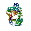 3agpS S: Starting model for refinement |
|---|---|
| Similar structure data |


-Links
 PDBj
PDBj


- Assembly
| Deposited unit | 
| ||||||||
|---|---|---|---|---|---|---|---|---|---|
| 1 | 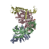
| ||||||||
| 2 | 
| ||||||||
| Unit cell |
|
-Components
-Elongation factor ... , 2 types, 4 molecules AEBF
| #1: Protein | Mass: 30332.795 Da / Num. of mol.: 2 Source method: isolated from a genetically manipulated source Source: (gene. exp.) #2: Protein | Mass: 43209.270 Da / Num. of mol.: 2 Source method: isolated from a genetically manipulated source Source: (gene. exp.) |
|---|
-Protein , 2 types, 4 molecules CGDH
| #3: Protein | Mass: 66305.039 Da / Num. of mol.: 2 Source method: isolated from a genetically manipulated source Source: (gene. exp.) Enterobacteria phage Qbeta (virus) / Production host: #4: Protein | Mass: 31340.545 Da / Num. of mol.: 2 / Fragment: UNP residues 1-273 Source method: isolated from a genetically manipulated source Source: (gene. exp.) |
|---|
-Non-polymers , 2 types, 257 molecules 

| #5: Chemical | ChemComp-SO4 /  Mass: 96.063 Da / Num. of mol.: 4 / Source method: obtained synthetically / Formula: SO4 Mass: 96.063 Da / Num. of mol.: 4 / Source method: obtained synthetically / Formula: SO4#6: Water | ChemComp-HOH / | Mass: 18.015 Da / Num. of mol.: 253 / Source method: isolated from a natural source / Formula: H2O |
|---|
-Experimental details
-Experiment
| Experiment | Method: X-RAY DIFFRACTION / Number of used crystals: 1 |
|---|
- Sample preparation
| Crystal | Density Matthews: 2.76 Å3/Da / Density % sol: 55.48 % |
|---|---|
| Crystal grow | Temperature: 293 K / Method: vapor diffusion, sitting drop / pH: 8 Details: 0.1M Tris, 0.2M lithium sulfate, 20% PEG3350, pH 8.0, VAPOR DIFFUSION, SITTING DROP, temperature 293K |
-Data collection
| Diffraction | Mean temperature: 95 K |
|---|---|
| Diffraction source | Source: SYNCHROTRON / Site: Photon Factory  / Beamline: BL-17A / Wavelength: 0.98 Å / Beamline: BL-17A / Wavelength: 0.98 Å |
| Detector | Type: ADSC QUANTUM 270 / Detector: CCD / Date: Feb 5, 2014 |
| Radiation | Protocol: SINGLE WAVELENGTH / Monochromatic (M) / Laue (L): M / Scattering type: x-ray |
| Radiation wavelength | Wavelength: 0.98 Å / Relative weight: 1 |
| Reflection | Resolution: 2.9→50 Å / Num. obs: 84212 / Biso Wilson estimate: 43.49 Å2 |
| Reflection shell | Resolution: 2.9→2.95 Å / Redundancy: 2.3 % / Mean I/σ(I) obs: 2.58 / % possible all: 86.2 |
- Processing
| Software |
| |||||||||||||||||||||||||||||||||||||||||||||||||||||||||||||||||||||||||||||||||||||||||||||||||||||||||||||||||||||||||||||||||||||||||||||||||||||||||||||||||||||||||||||||||||||||||||||||||||||||||||||||||||||||||
|---|---|---|---|---|---|---|---|---|---|---|---|---|---|---|---|---|---|---|---|---|---|---|---|---|---|---|---|---|---|---|---|---|---|---|---|---|---|---|---|---|---|---|---|---|---|---|---|---|---|---|---|---|---|---|---|---|---|---|---|---|---|---|---|---|---|---|---|---|---|---|---|---|---|---|---|---|---|---|---|---|---|---|---|---|---|---|---|---|---|---|---|---|---|---|---|---|---|---|---|---|---|---|---|---|---|---|---|---|---|---|---|---|---|---|---|---|---|---|---|---|---|---|---|---|---|---|---|---|---|---|---|---|---|---|---|---|---|---|---|---|---|---|---|---|---|---|---|---|---|---|---|---|---|---|---|---|---|---|---|---|---|---|---|---|---|---|---|---|---|---|---|---|---|---|---|---|---|---|---|---|---|---|---|---|---|---|---|---|---|---|---|---|---|---|---|---|---|---|---|---|---|---|---|---|---|---|---|---|---|---|---|---|---|---|---|---|---|---|
| Refinement | Method to determine structure: MOLECULAR REPLACEMENT Starting model: 3AGP Resolution: 2.9→19.982 Å / FOM work R set: 0.7413 / SU ML: 0.6 / σ(F): 1.51 / Phase error: 32.5 / Stereochemistry target values: ML
| |||||||||||||||||||||||||||||||||||||||||||||||||||||||||||||||||||||||||||||||||||||||||||||||||||||||||||||||||||||||||||||||||||||||||||||||||||||||||||||||||||||||||||||||||||||||||||||||||||||||||||||||||||||||||
| Solvent computation | Shrinkage radii: 0.9 Å / VDW probe radii: 1.11 Å / Solvent model: FLAT BULK SOLVENT MODEL | |||||||||||||||||||||||||||||||||||||||||||||||||||||||||||||||||||||||||||||||||||||||||||||||||||||||||||||||||||||||||||||||||||||||||||||||||||||||||||||||||||||||||||||||||||||||||||||||||||||||||||||||||||||||||
| Displacement parameters | Biso max: 175.49 Å2 / Biso mean: 63.95 Å2 / Biso min: 6.88 Å2 | |||||||||||||||||||||||||||||||||||||||||||||||||||||||||||||||||||||||||||||||||||||||||||||||||||||||||||||||||||||||||||||||||||||||||||||||||||||||||||||||||||||||||||||||||||||||||||||||||||||||||||||||||||||||||
| Refinement step | Cycle: LAST / Resolution: 2.9→19.982 Å
| |||||||||||||||||||||||||||||||||||||||||||||||||||||||||||||||||||||||||||||||||||||||||||||||||||||||||||||||||||||||||||||||||||||||||||||||||||||||||||||||||||||||||||||||||||||||||||||||||||||||||||||||||||||||||
| Refine LS restraints |
| |||||||||||||||||||||||||||||||||||||||||||||||||||||||||||||||||||||||||||||||||||||||||||||||||||||||||||||||||||||||||||||||||||||||||||||||||||||||||||||||||||||||||||||||||||||||||||||||||||||||||||||||||||||||||
| LS refinement shell | Refine-ID: X-RAY DIFFRACTION / Total num. of bins used: 30
|
