 Movie
Movie Controller


+ Open data

- Basic information
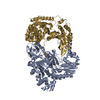
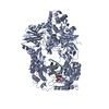
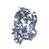
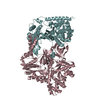
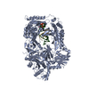
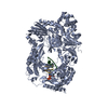
| Entry | Database: PDB / ID: 3agq | ||||||
|---|---|---|---|---|---|---|---|
| Title | Structure of viral polymerase form II | ||||||
 Components Components | Elongation factor Ts, Elongation factor Tu 1, LINKER, Q beta replicase | ||||||
 Keywords Keywords | TRANSLATION / TRANSFERASE / RNA polymerase / replicase | ||||||
| Function / homology |  Function and homology information Function and homology informationguanyl-nucleotide exchange factor complex / protein-synthesizing GTPase / guanosine tetraphosphate binding / translation elongation factor activity / RNA-directed RNA polymerase / response to antibiotic / viral RNA genome replication / nucleotide binding / RNA-directed RNA polymerase activity / GTPase activity ...guanyl-nucleotide exchange factor complex / protein-synthesizing GTPase / guanosine tetraphosphate binding / translation elongation factor activity / RNA-directed RNA polymerase / response to antibiotic / viral RNA genome replication / nucleotide binding / RNA-directed RNA polymerase activity / GTPase activity / GTP binding / magnesium ion binding / RNA binding / metal ion binding / plasma membrane / cytosol / cytoplasm Similarity search - Function | ||||||
| Biological species |  synthetic construct (others)  Escherichia phage Qbeta (virus) Escherichia phage Qbeta (virus) | ||||||
| Method |  X-RAY DIFFRACTION / SYNCHROTRON / MOLECULAR REPLACEMENT / Resolution: 3.22 Å X-RAY DIFFRACTION / SYNCHROTRON / MOLECULAR REPLACEMENT / Resolution: 3.22 Å | ||||||
 Authors Authors | Takeshita, D. / Tomita, K. | ||||||
 Citation Citation | Journal: Proc.Natl.Acad.Sci.USA / Year: 2010 Title: Assembly of Q{beta} viral RNA polymerase with host translational elongation factors EF-Tu and -Ts Authors: Takeshita, D. / Tomita, K. | ||||||
| History |
|
- Structure visualization
| Structure viewer | Molecule: MolmilJmol/JSmol |
|---|
- Downloads & links
-Download
| PDBx/mmCIF format | 3agq.cif.gz | 243.6 KB | Display | PDBx/mmCIF format |
|---|---|---|---|---|
| PDB format | pdb3agq.ent.gz | 191 KB | Display | PDB format |
| PDBx/mmJSON format | 3agq.json.gz | Tree view | PDBx/mmJSON format | |
| Others |  Other downloads Other downloads |
-Validation report
| Arichive directory | https://data.pdbj.org/pub/pdb/validation_reports/ag/3agqftp://data.pdbj.org/pub/pdb/validation_reports/ag/3agq | HTTPS FTP |
|---|
-Related structure data
| Related structure data |  3agpSC S: Starting model for refinement C: citing same article ( |
|---|---|
| Similar structure data |


-Links
 PDBj
PDBj


- Assembly
| Deposited unit | 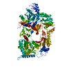
| ||||||||
|---|---|---|---|---|---|---|---|---|---|
| 1 |
| ||||||||
| 2 | 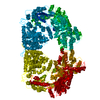
| ||||||||
| Unit cell |
|
-Components
| #1: Protein | Mass: 141417.688 Da / Num. of mol.: 1 Source method: isolated from a genetically manipulated source Source: (gene. exp.) Escherichia phage Qbeta (virus)Plasmid: pBAD33 / Production host: References: UniProt: P0A6P3, UniProt: P0A6N3, UniProt: Q8LTE0 |
|---|---|
| #2: Chemical | ChemComp-MG /   Mass: 24.305 Da / Num. of mol.: 1 / Source method: obtained synthetically / Formula: Mg Mass: 24.305 Da / Num. of mol.: 1 / Source method: obtained synthetically / Formula: Mg |
| #3: Water | ChemComp-HOH /  Mass: 18.015 Da / Num. of mol.: 4 / Source method: isolated from a natural source / Formula: H2O Mass: 18.015 Da / Num. of mol.: 4 / Source method: isolated from a natural source / Formula: H2O |
-Experimental details
-Experiment
| Experiment | Method: X-RAY DIFFRACTION / Number of used crystals: 1 |
|---|
- Sample preparation
| Crystal | Density Matthews: 3.16 Å3/Da / Density % sol: 61.07 % |
|---|---|
| Crystal grow | Temperature: 293 K / Method: vapor diffusion, sitting drop / pH: 5.6 Details: 32% PEG400, 0.2M magnesium acetate, 0.1M HEPES, pH 5.6, VAPOR DIFFUSION, SITTING DROP, temperature 293K |
-Data collection
| Diffraction | Mean temperature: 95 K |
|---|---|
| Diffraction source | Source: SYNCHROTRON / Site: Photon Factory  / Beamline: BL-17A / Wavelength: 0.98 Å / Beamline: BL-17A / Wavelength: 0.98 Å |
| Detector | Type: ADSC QUANTUM 210r / Detector: CCD / Date: Nov 11, 2009 |
| Radiation | Protocol: SINGLE WAVELENGTH / Monochromatic (M) / Laue (L): M / Scattering type: x-ray |
| Radiation wavelength | Wavelength: 0.98 Å / Relative weight: 1 |
| Reflection | Resolution: 3.2→30 Å / Num. obs: 28658 / % possible obs: 98.5 % / Redundancy: 8.4 % / Rmerge(I) obs: 0.107 |
| Reflection shell | Resolution: 3.2→3.26 Å / Redundancy: 4.5 % / Rmerge(I) obs: 0.341 / Num. unique all: 1396 / % possible all: 98.1 |
- Processing
| Software |
| ||||||||||||||||
|---|---|---|---|---|---|---|---|---|---|---|---|---|---|---|---|---|---|
| Refinement | Method to determine structure: MOLECULAR REPLACEMENT Starting model: 3AGP Resolution: 3.22→20 Å
| ||||||||||||||||
| Refinement step | Cycle: LAST / Resolution: 3.22→20 Å
| ||||||||||||||||
| Refine LS restraints |
| ||||||||||||||||
| LS refinement shell | Resolution: 3.2213→3.2414 Å / Rfactor Rwork: 0.476 |
