 Movie
Movie Controller

[English] 日本語
 Yorodumi
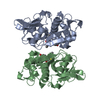
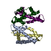
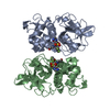
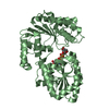
- PDB-7bgm: Crystal structure of MtHISN2, a bifunctional enzyme from the hist... -
+ Open data

- Basic information
| Entry | Database: PDB / ID: 7bgm | ||||||
|---|---|---|---|---|---|---|---|
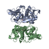
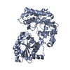
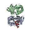
| Title | Crystal structure of MtHISN2, a bifunctional enzyme from the histidine biosynthetic pathway | ||||||
 Components Components | Phosphoribosyl-AMP cyclohydrolase | ||||||
 Keywords Keywords | HYDROLASE / Histidine / Pyrophosphohydrolase / Cyclohydrolase / Bifunctional / PRA-CH / PRA-PH | ||||||
| Function / homology |  Function and homology information Function and homology informationphosphoribosyl-AMP cyclohydrolase activity / phosphoribosyl-ATP diphosphatase activity / L-histidine biosynthetic process / ATP binding / metal ion binding Similarity search - Function | ||||||
| Biological species |  | ||||||
| Method |  X-RAY DIFFRACTION / SYNCHROTRON / SAD / Resolution: 1.6 Å X-RAY DIFFRACTION / SYNCHROTRON / SAD / Resolution: 1.6 Å | ||||||
 Authors Authors | Witek, W. / Ruszkowski, M. | ||||||
| Funding support |  Poland, 1items Poland, 1items
| ||||||
 Citation Citation | Journal: Sci Rep / Year: 2021 Title: Structural and mechanistic insights into the bifunctional HISN2 enzyme catalyzing the second and third steps of histidine biosynthesis in plants. Authors: Witek, W. / Sliwiak, J. / Ruszkowski, M. | ||||||
| History |
|
- Structure visualization
| Structure viewer | Molecule: MolmilJmol/JSmol |
|---|
- Downloads & links
-Download
| PDBx/mmCIF format | 7bgm.cif.gz | 233.6 KB | Display | PDBx/mmCIF format |
|---|---|---|---|---|
| PDB format | pdb7bgm.ent.gz | 157.8 KB | Display | PDB format |
| PDBx/mmJSON format | 7bgm.json.gz | Tree view | PDBx/mmJSON format | |
| Others |  Other downloads Other downloads |
-Validation report
| Arichive directory | https://data.pdbj.org/pub/pdb/validation_reports/bg/7bgmftp://data.pdbj.org/pub/pdb/validation_reports/bg/7bgm | HTTPS FTP |
|---|
-Related structure data
| Related structure data |  7bgnC C: citing same article ( |
|---|---|
| Similar structure data | |
| Experimental dataset #1 | Data reference: 10.18150/WRT4WT / Data set type: diffraction image data |


-Links
 PDBj
PDBj- Assembly
| Deposited unit | 
| ||||||||||||
|---|---|---|---|---|---|---|---|---|---|---|---|---|---|
| 1 |
| ||||||||||||
| Unit cell |
| ||||||||||||
| Components on special symmetry positions |
|
-Components
| #1: Protein | Mass: 26692.064 Da / Num. of mol.: 2 Source method: isolated from a genetically manipulated source Details: Without a signal peptide (residues 1-48) / Source: (gene. exp.) Production host:  References: UniProt: A0A072U2X9, phosphoribosyl-AMP cyclohydrolase, phosphoribosyl-ATP diphosphatase #2: Chemical | ChemComp-ZN /   Mass: 65.409 Da / Num. of mol.: 9 / Source method: obtained synthetically / Formula: Zn Mass: 65.409 Da / Num. of mol.: 9 / Source method: obtained synthetically / Formula: Zn#3: Water | ChemComp-HOH / |  Mass: 18.015 Da / Num. of mol.: 356 / Source method: isolated from a natural source / Formula: H2O Mass: 18.015 Da / Num. of mol.: 356 / Source method: isolated from a natural source / Formula: H2OHas ligand of interest | N | |
|---|
-Experimental details
-Experiment
| Experiment | Method: X-RAY DIFFRACTION / Number of used crystals: 1 |
|---|
- Sample preparation
| Crystal | Density Matthews: 3.27 Å3/Da / Density % sol: 62.34 % |
|---|---|
| Crystal grow | Temperature: 291 K / Method: vapor diffusion, sitting drop / pH: 6.5 Details: 0.12 M Alcohols (0.2M 1,6-Hexanediol; 0.2M 1-Butanol 0.2M 1,2-Propanediol; 0.2M 2-Propanol; 0.2M 1,4-Butanediol; 0.2M 1,3-Propanediol) 0.1 M Buffer System 1 , pH 6.5 (Imidazole; MES-acid) ...Details: 0.12 M Alcohols (0.2M 1,6-Hexanediol; 0.2M 1-Butanol 0.2M 1,2-Propanediol; 0.2M 2-Propanol; 0.2M 1,4-Butanediol; 0.2M 1,3-Propanediol) 0.1 M Buffer System 1 , pH 6.5 (Imidazole; MES-acid) 30% Precipitant Mix 1 (20% v/v PEG 500* MME; 10 % w/v PEG 20000. Cryoprotection: 20% ethylene glycol. |
-Data collection
| Diffraction | Mean temperature: 100 K / Serial crystal experiment: N |
|---|---|
| Diffraction source | Source: SYNCHROTRON / Site: APS  / Beamline: 19-ID / Wavelength: 0.9793 Å / Beamline: 19-ID / Wavelength: 0.9793 Å |
| Detector | Type: DECTRIS PILATUS3 6M / Detector: PIXEL / Date: Dec 14, 2018 |
| Radiation | Monochromator: Si(111) / Protocol: SINGLE WAVELENGTH / Monochromatic (M) / Laue (L): M / Scattering type: x-ray |
| Radiation wavelength | Wavelength: 0.9793 Å / Relative weight: 1 |
| Reflection | Resolution: 1.6→80 Å / Num. obs: 79751 / % possible obs: 98.5 % / Redundancy: 3.7 % / Biso Wilson estimate: 25.3 Å2 / CC1/2: 0.99 / Rmerge(I) obs: 0.045 / Net I/σ(I): 14.6 |
| Reflection shell | Resolution: 1.6→1.7 Å / Redundancy: 3.8 % / Rmerge(I) obs: 0.62 / Mean I/σ(I) obs: 1.9 / Num. unique obs: 12569 / CC1/2: 0.69 / % possible all: 96.3 |
- Processing
| Software |
| ||||||||||||||||||||||||||||||||||||||||||||||||||||||||
|---|---|---|---|---|---|---|---|---|---|---|---|---|---|---|---|---|---|---|---|---|---|---|---|---|---|---|---|---|---|---|---|---|---|---|---|---|---|---|---|---|---|---|---|---|---|---|---|---|---|---|---|---|---|---|---|---|---|
| Refinement | Method to determine structure: SAD / Resolution: 1.6→42.93 Å / SU ML: 0.155 / Cross valid method: FREE R-VALUE / Phase error: 18.9436 Stereochemistry target values: GeoStd + Monomer Library + CDL v1.2
| ||||||||||||||||||||||||||||||||||||||||||||||||||||||||
| Solvent computation | Shrinkage radii: 0.9 Å / VDW probe radii: 1.11 Å / Solvent model: FLAT BULK SOLVENT MODEL | ||||||||||||||||||||||||||||||||||||||||||||||||||||||||
| Displacement parameters | Biso mean: 38.58 Å2 | ||||||||||||||||||||||||||||||||||||||||||||||||||||||||
| Refinement step | Cycle: LAST / Resolution: 1.6→42.93 Å
| ||||||||||||||||||||||||||||||||||||||||||||||||||||||||
| Refine LS restraints |
| ||||||||||||||||||||||||||||||||||||||||||||||||||||||||
| LS refinement shell |
|
