 Movie
Movie Controller

[English] 日本語
 Yorodumi
- PDB-3mha: Crystal structure of LprG from Mycobacterium tuberculosis bound to PIM -
+ Open data

- Basic information
| Entry | Database: PDB / ID: 3mha | ||||||
|---|---|---|---|---|---|---|---|
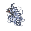
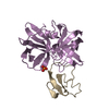
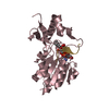
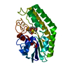
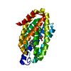
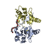
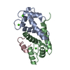
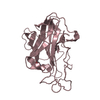
| Title | Crystal structure of LprG from Mycobacterium tuberculosis bound to PIM | ||||||
 Components Components | Lipoprotein lprG | ||||||
 Keywords Keywords | LIPID BINDING PROTEIN / Lipoprotein / LprG / glycolipid binding protein / Structural Genomics / TB Structural Genomics Consortium / TBSGC | ||||||
| Function / homology |  Function and homology information Function and homology informationbacterial extracellular vesicle / glycolipid binding / lipid transport / phosphatidylinositol binding / peptidoglycan-based cell wall / Prevention of phagosomal-lysosomal fusion / response to antibiotic / lipid binding / cell surface / extracellular region ...bacterial extracellular vesicle / glycolipid binding / lipid transport / phosphatidylinositol binding / peptidoglycan-based cell wall / Prevention of phagosomal-lysosomal fusion / response to antibiotic / lipid binding / cell surface / extracellular region / plasma membrane / cytosol Similarity search - Function | ||||||
| Biological species |   Mycobacterium tuberculosis (bacteria) Mycobacterium tuberculosis (bacteria) | ||||||
| Method |  X-RAY DIFFRACTION / SYNCHROTRON / MOLECULAR REPLACEMENT / Resolution: 1.85 Å X-RAY DIFFRACTION / SYNCHROTRON / MOLECULAR REPLACEMENT / Resolution: 1.85 Å | ||||||
 Authors Authors | Tsai, H.-C. / Sacchettini, J.C. / TB Structural Genomics Consortium (TBSGC) | ||||||
 Citation Citation | Journal: Nat.Struct.Mol.Biol. / Year: 2010 Title: Mycobacterium tuberculosis lipoprotein LprG (Rv1411c) binds triacylated glycolipid agonists of Toll-like receptor 2. Authors: Drage, M.G. / Tsai, H.C. / Pecora, N.D. / Cheng, T.Y. / Arida, A.R. / Shukla, S. / Rojas, R.E. / Seshadri, C. / Moody, D.B. / Boom, W.H. / Sacchettini, J.C. / Harding, C.V. | ||||||
| History |
|
- Structure visualization
| Structure viewer | Molecule: MolmilJmol/JSmol |
|---|
- Downloads & links
-Download
| PDBx/mmCIF format | 3mha.cif.gz | 92.2 KB | Display | PDBx/mmCIF format |
|---|---|---|---|---|
| PDB format | pdb3mha.ent.gz | 68 KB | Display | PDB format |
| PDBx/mmJSON format | 3mha.json.gz | Tree view | PDBx/mmJSON format | |
| Others |  Other downloads Other downloads |
-Validation report
| Arichive directory | https://data.pdbj.org/pub/pdb/validation_reports/mh/3mhaftp://data.pdbj.org/pub/pdb/validation_reports/mh/3mha | HTTPS FTP |
|---|
-Related structure data
| Related structure data |  3mh8SC 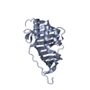 3mh9C S: Starting model for refinement C: citing same article ( |
|---|---|
| Similar structure data | |
| Other databases |


-Links
 PDBj
PDBj


- Assembly
| Deposited unit | 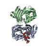
| ||||||||
|---|---|---|---|---|---|---|---|---|---|
| 1 | 
| ||||||||
| 2 | 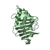
| ||||||||
| Unit cell |
|
-Components
| #1: Protein | Mass: 21464.941 Da / Num. of mol.: 2 Source method: isolated from a genetically manipulated source Source: (gene. exp.) Mycobacterium tuberculosis (bacteria) / Strain: H37Rv / Gene: lpp-27, lprG, MT1455, MTCY21B4.28c, Rv1411c / Plasmid: pET30b / Production host: #2: Chemical | ChemComp-Z69 / ( |   Mass: 1421.849 Da / Num. of mol.: 1 / Source method: obtained synthetically / Formula: C72H141O24P Mass: 1421.849 Da / Num. of mol.: 1 / Source method: obtained synthetically / Formula: C72H141O24P#3: Water | ChemComp-HOH / |  Mass: 18.015 Da / Num. of mol.: 175 / Source method: isolated from a natural source / Formula: H2O Mass: 18.015 Da / Num. of mol.: 175 / Source method: isolated from a natural source / Formula: H2O |
|---|
-Experimental details
-Experiment
| Experiment | Method: X-RAY DIFFRACTION / Number of used crystals: 1 |
|---|
- Sample preparation
| Crystal | Density Matthews: 2.33 Å3/Da / Density % sol: 47.12 % |
|---|---|
| Crystal grow | Temperature: 289 K / Method: vapor diffusion, hanging drop / pH: 4.5 Details: 100mM Sodium acetate trihydrate, 25% PEG3350, pH 4.5, VAPOR DIFFUSION, HANGING DROP, temperature 289K |
-Data collection
| Diffraction | Mean temperature: 120 K |
|---|---|
| Diffraction source | Source: SYNCHROTRON / Site: APS  / Beamline: 23-ID-B / Wavelength: 0.9795 Å / Beamline: 23-ID-B / Wavelength: 0.9795 Å |
| Detector | Type: MARMOSAIC 300 mm CCD / Detector: CCD / Date: Feb 14, 2009 |
| Radiation | Monochromator: mirror / Protocol: SINGLE WAVELENGTH / Monochromatic (M) / Laue (L): M / Scattering type: x-ray |
| Radiation wavelength | Wavelength: 0.9795 Å / Relative weight: 1 |
| Reflection | Resolution: 1.73→50 Å / Num. all: 41304 / Num. obs: 35778 / % possible obs: 86.6 % / Observed criterion σ(F): 1 / Observed criterion σ(I): 1 / Redundancy: 5 % / Rmerge(I) obs: 0.052 / Rsym value: 0.061 / Net I/σ(I): 4.7 |
| Reflection shell | Resolution: 1.85→1.92 Å / Redundancy: 2.8 % / Rmerge(I) obs: 0.624 / Mean I/σ(I) obs: 5.5 / Num. unique all: 2021 / Rsym value: 0.721 / % possible all: 82.2 |
- Processing
| Software | Name: PHENIX / Version: (phenix.refine: 1.6_289) / Classification: refinement | ||||||||||||||||||||||||||||||||||||||||||||||||||||||||||||||||||||||||||||||||||||||||||||||||||
|---|---|---|---|---|---|---|---|---|---|---|---|---|---|---|---|---|---|---|---|---|---|---|---|---|---|---|---|---|---|---|---|---|---|---|---|---|---|---|---|---|---|---|---|---|---|---|---|---|---|---|---|---|---|---|---|---|---|---|---|---|---|---|---|---|---|---|---|---|---|---|---|---|---|---|---|---|---|---|---|---|---|---|---|---|---|---|---|---|---|---|---|---|---|---|---|---|---|---|---|
| Refinement | Method to determine structure: MOLECULAR REPLACEMENT Starting model: 3MH8 Resolution: 1.85→32.047 Å / SU ML: 0.25 / σ(F): 1.35 / Stereochemistry target values: ML
| ||||||||||||||||||||||||||||||||||||||||||||||||||||||||||||||||||||||||||||||||||||||||||||||||||
| Solvent computation | Shrinkage radii: 0.9 Å / VDW probe radii: 1.11 Å / Solvent model: FLAT BULK SOLVENT MODEL / Bsol: 41.697 Å2 / ksol: 0.332 e/Å3 | ||||||||||||||||||||||||||||||||||||||||||||||||||||||||||||||||||||||||||||||||||||||||||||||||||
| Refinement step | Cycle: LAST / Resolution: 1.85→32.047 Å
| ||||||||||||||||||||||||||||||||||||||||||||||||||||||||||||||||||||||||||||||||||||||||||||||||||
| Refine LS restraints |
| ||||||||||||||||||||||||||||||||||||||||||||||||||||||||||||||||||||||||||||||||||||||||||||||||||
| LS refinement shell |
|
