 Movie
Movie Controller

[English] 日本語
 Yorodumi
- PDB-4tw3: Insights into Substrate and Metal Binding from the Crystal Struct... -
+ Open data

- Basic information
| Entry | Database: PDB / ID: 4tw3 | ||||||
|---|---|---|---|---|---|---|---|
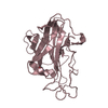
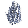
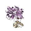
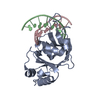
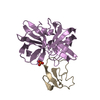
| Title | Insights into Substrate and Metal Binding from the Crystal Structure of Cyanobacterial Aldehyde Deformylating Oxygenase with Substrate Bound | ||||||
 Components Components | Aldehyde decarbonylase | ||||||
 Keywords Keywords | OXIDOREDUCTASE / non-heme di-iron protein / hydrocarbon production / alpha-helix | ||||||
| Function / homology |  Function and homology information Function and homology informationaldehyde oxygenase (deformylating) activity / aldehyde oxygenase (deformylating) / transition metal ion binding Similarity search - Function | ||||||
| Biological species |  Prochlorococcus marinus (bacteria) Prochlorococcus marinus (bacteria) | ||||||
| Method |  X-RAY DIFFRACTION / SYNCHROTRON / MOLECULAR REPLACEMENT / Resolution: 1.6 Å X-RAY DIFFRACTION / SYNCHROTRON / MOLECULAR REPLACEMENT / Resolution: 1.6 Å | ||||||
 Authors Authors | Buer, B.C. / Paul, B. / Das, D. / Stuckey, J.A. / Marsh, E.N.G. | ||||||
 Citation Citation | Journal: Acs Chem.Biol. / Year: 2014 Title: Insights into substrate and metal binding from the crystal structure of cyanobacterial aldehyde deformylating oxygenase with substrate bound. Authors: Buer, B.C. / Paul, B. / Das, D. / Stuckey, J.A. / Marsh, E.N. | ||||||
| History |
|
- Structure visualization
| Structure viewer | Molecule: MolmilJmol/JSmol |
|---|
- Downloads & links
-Download
| PDBx/mmCIF format | 4tw3.cif.gz | 112.3 KB | Display | PDBx/mmCIF format |
|---|---|---|---|---|
| PDB format | pdb4tw3.ent.gz | 83.4 KB | Display | PDB format |
| PDBx/mmJSON format | 4tw3.json.gz | Tree view | PDBx/mmJSON format | |
| Others |  Other downloads Other downloads |
-Validation report
| Arichive directory | https://data.pdbj.org/pub/pdb/validation_reports/tw/4tw3ftp://data.pdbj.org/pub/pdb/validation_reports/tw/4tw3 | HTTPS FTP |
|---|
-Related structure data
| Related structure data | 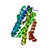 4pg0C 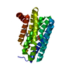 4pg1C 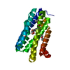 4pgiC 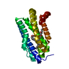 4pgkC  2oc5S S: Starting model for refinement C: citing same article ( |
|---|---|
| Similar structure data |


-Links
 PDBj
PDBj


- Assembly
| Deposited unit | 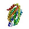
| ||||||||
|---|---|---|---|---|---|---|---|---|---|
| 1 |
| ||||||||
| Unit cell |
|
-Components
| #1: Protein | Mass: 25191.684 Da / Num. of mol.: 1 Source method: isolated from a genetically manipulated source Source: (gene. exp.) Prochlorococcus marinus (bacteria) / Strain: MIT 9313 / Gene: PMT_1231 / Production host: References: UniProt: Q7V6D4, aldehyde oxygenase (deformylating) | ||||||
|---|---|---|---|---|---|---|---|
| #2: Chemical |   Mass: 55.845 Da / Num. of mol.: 2 / Source method: obtained synthetically / Formula: Fe Mass: 55.845 Da / Num. of mol.: 2 / Source method: obtained synthetically / Formula: Fe#3: Chemical | ChemComp-STE / |   Mass: 284.477 Da / Num. of mol.: 1 / Source method: obtained synthetically / Formula: C18H36O2 Mass: 284.477 Da / Num. of mol.: 1 / Source method: obtained synthetically / Formula: C18H36O2#4: Chemical | ChemComp-EDO / |   Mass: 62.068 Da / Num. of mol.: 1 / Source method: obtained synthetically / Formula: C2H6O2 Mass: 62.068 Da / Num. of mol.: 1 / Source method: obtained synthetically / Formula: C2H6O2#5: Water | ChemComp-HOH / |  Mass: 18.015 Da / Num. of mol.: 293 / Source method: isolated from a natural source / Formula: H2O Mass: 18.015 Da / Num. of mol.: 293 / Source method: isolated from a natural source / Formula: H2O |
-Experimental details
-Experiment
| Experiment | Method: X-RAY DIFFRACTION |
|---|
- Sample preparation
| Crystal | Density Matthews: 3.46 Å3/Da / Density % sol: 64.46 % |
|---|---|
| Crystal grow | Temperature: 277 K / Method: vapor diffusion, hanging drop / Details: 25% PEG 1000, 0.1 M HEPES / PH range: 7.5 |
-Data collection
| Diffraction | Mean temperature: 100 K |
|---|---|
| Diffraction source | Source: SYNCHROTRON / Site: APS  / Beamline: 21-ID-F / Wavelength: 0.97872 Å / Beamline: 21-ID-F / Wavelength: 0.97872 Å |
| Detector | Type: MARMOSAIC 225 mm CCD / Detector: CCD / Date: Apr 12, 2013 |
| Radiation | Protocol: SINGLE WAVELENGTH / Monochromatic (M) / Laue (L): M / Scattering type: x-ray |
| Radiation wavelength | Wavelength: 0.97872 Å / Relative weight: 1 |
| Reflection | Resolution: 1.6→38.61 Å / Num. obs: 47171 / % possible obs: 100 % / Redundancy: 14.4 % / Biso Wilson estimate: 20.98 Å2 / Net I/σ(I): 20 |
| Reflection shell | Resolution: 1.6→1.63 Å / Redundancy: 14.3 % / Mean I/σ(I) obs: 3 / % possible all: 100 |
- Processing
| Software | Name: BUSTER / Version: 2.10.0 / Classification: refinement | ||||||||||||||||||||||||||||||||||||||||||||||||||||||||||||||||||||||||||||||||||||||||||||||||||||||||||||||||||
|---|---|---|---|---|---|---|---|---|---|---|---|---|---|---|---|---|---|---|---|---|---|---|---|---|---|---|---|---|---|---|---|---|---|---|---|---|---|---|---|---|---|---|---|---|---|---|---|---|---|---|---|---|---|---|---|---|---|---|---|---|---|---|---|---|---|---|---|---|---|---|---|---|---|---|---|---|---|---|---|---|---|---|---|---|---|---|---|---|---|---|---|---|---|---|---|---|---|---|---|---|---|---|---|---|---|---|---|---|---|---|---|---|---|---|---|
| Refinement | Method to determine structure: MOLECULAR REPLACEMENT Starting model: 2OC5 Resolution: 1.6→38.61 Å / Cor.coef. Fo:Fc: 0.9577 / Cor.coef. Fo:Fc free: 0.9559 / SU R Cruickshank DPI: 0.066 / Cross valid method: THROUGHOUT / σ(F): 0 / SU R Blow DPI: 0.071 / SU Rfree Blow DPI: 0.068 / SU Rfree Cruickshank DPI: 0.064
| ||||||||||||||||||||||||||||||||||||||||||||||||||||||||||||||||||||||||||||||||||||||||||||||||||||||||||||||||||
| Displacement parameters | Biso mean: 22.29 Å2
| ||||||||||||||||||||||||||||||||||||||||||||||||||||||||||||||||||||||||||||||||||||||||||||||||||||||||||||||||||
| Refine analyze | Luzzati coordinate error obs: 0.172 Å | ||||||||||||||||||||||||||||||||||||||||||||||||||||||||||||||||||||||||||||||||||||||||||||||||||||||||||||||||||
| Refinement step | Cycle: 1 / Resolution: 1.6→38.61 Å
| ||||||||||||||||||||||||||||||||||||||||||||||||||||||||||||||||||||||||||||||||||||||||||||||||||||||||||||||||||
| Refine LS restraints |
| ||||||||||||||||||||||||||||||||||||||||||||||||||||||||||||||||||||||||||||||||||||||||||||||||||||||||||||||||||
| LS refinement shell | Resolution: 1.6→1.64 Å / Total num. of bins used: 20
| ||||||||||||||||||||||||||||||||||||||||||||||||||||||||||||||||||||||||||||||||||||||||||||||||||||||||||||||||||
| Refinement TLS params. | Method: refined / Origin x: -20.4648 Å / Origin y: 1.8418 Å / Origin z: -9.5515 Å
| ||||||||||||||||||||||||||||||||||||||||||||||||||||||||||||||||||||||||||||||||||||||||||||||||||||||||||||||||||
| Refinement TLS group | Selection details: { A|21 - A|242 } |
