 Movie
Movie Controller

[English] 日本語
 Yorodumi
- PDB-5k52: Crystal structures of aldehyde deformylating oxygenase from Limno... -
+ Open data

- Basic information
| Entry | Database: PDB / ID: 5k52 | ||||||
|---|---|---|---|---|---|---|---|
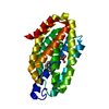
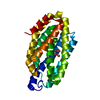
| Title | Crystal structures of aldehyde deformylating oxygenase from Limnothrix sp. KNUA012 | ||||||
 Components Components | Aldehyde decarbonylase | ||||||
 Keywords Keywords | METAL BINDING PROTEIN / ferritin-like di-iron protein | ||||||
| Function / homology |  Function and homology information Function and homology informationaldehyde oxygenase (deformylating) activity / aldehyde oxygenase (deformylating) / transition metal ion binding Similarity search - Function | ||||||
| Biological species |  Limnothrix sp. KNUA012 (bacteria) Limnothrix sp. KNUA012 (bacteria) | ||||||
| Method |  X-RAY DIFFRACTION / SYNCHROTRON / MOLECULAR REPLACEMENT / Resolution: 2.4 Å X-RAY DIFFRACTION / SYNCHROTRON / MOLECULAR REPLACEMENT / Resolution: 2.4 Å | ||||||
 Authors Authors | Park, A.K. / Kim, H-.W. | ||||||
| Funding support |  Korea, Republic Of, 1items Korea, Republic Of, 1items
| ||||||
 Citation Citation | Journal: Biochem. Biophys. Res. Commun. / Year: 2016 Title: Crystal structures of aldehyde deformylating oxygenase from Limnothrix sp. KNUA012 and Oscillatoria sp. KNUA011. Authors: Park, A.K. / Kim, I.S. / Jeon, B.W. / Roh, S.J. / Ryu, M.Y. / Baek, H.R. / Jo, S.W. / Kim, Y.S. / Park, H. / Lee, J.H. / Yoon, H.S. / Kim, H.W. | ||||||
| History |
|
- Structure visualization
| Structure viewer | Molecule: MolmilJmol/JSmol |
|---|
- Downloads & links
-Download
| PDBx/mmCIF format | 5k52.cif.gz | 188.7 KB | Display | PDBx/mmCIF format |
|---|---|---|---|---|
| PDB format | pdb5k52.ent.gz | 149.8 KB | Display | PDB format |
| PDBx/mmJSON format | 5k52.json.gz | Tree view | PDBx/mmJSON format | |
| Others |  Other downloads Other downloads |
-Validation report
| Arichive directory | https://data.pdbj.org/pub/pdb/validation_reports/k5/5k52ftp://data.pdbj.org/pub/pdb/validation_reports/k5/5k52 | HTTPS FTP |
|---|
-Related structure data
| Related structure data |  5k53C 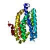 4quwS C: citing same article ( S: Starting model for refinement |
|---|---|
| Similar structure data |


-Links
 PDBj
PDBj


- Assembly
| Deposited unit | 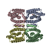
| ||||||||
|---|---|---|---|---|---|---|---|---|---|
| 1 | 
| ||||||||
| 2 | 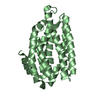
| ||||||||
| 3 | 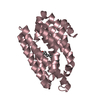
| ||||||||
| 4 | 
| ||||||||
| Unit cell |
|
-Components
| #1: Protein | Mass: 29417.166 Da / Num. of mol.: 4 Source method: isolated from a genetically manipulated source Source: (gene. exp.) Limnothrix sp. KNUA012 (bacteria) / Production host: References: UniProt: A0A193CDY9, aldehyde oxygenase (deformylating) #2: Chemical |   Mass: 268.478 Da / Num. of mol.: 2 / Source method: obtained synthetically / Formula: C18H36O Mass: 268.478 Da / Num. of mol.: 2 / Source method: obtained synthetically / Formula: C18H36O#3: Water | ChemComp-HOH / |  Mass: 18.015 Da / Num. of mol.: 118 / Source method: isolated from a natural source / Formula: H2O Mass: 18.015 Da / Num. of mol.: 118 / Source method: isolated from a natural source / Formula: H2OSequence details | A sequence reference for this protein does not currently exist. | |
|---|
-Experimental details
-Experiment
| Experiment | Method: X-RAY DIFFRACTION / Number of used crystals: 1 |
|---|
- Sample preparation
| Crystal | Density Matthews: 2.81 Å3/Da / Density % sol: 56.18 % |
|---|---|
| Crystal grow | Temperature: 296 K / Method: vapor diffusion, hanging drop / Details: Bis-Tris propane, sodium chloride |
-Data collection
| Diffraction | Mean temperature: 100 K | |||||||||||||||||||||||||||||||||||||||||||||||||||||||||||||||||||||||||||||||||||||||||||||||||||||||||||||||||||||||||||||||||||||||||||||||||||||||||||||||||||||||||||||||||||||||||||||
|---|---|---|---|---|---|---|---|---|---|---|---|---|---|---|---|---|---|---|---|---|---|---|---|---|---|---|---|---|---|---|---|---|---|---|---|---|---|---|---|---|---|---|---|---|---|---|---|---|---|---|---|---|---|---|---|---|---|---|---|---|---|---|---|---|---|---|---|---|---|---|---|---|---|---|---|---|---|---|---|---|---|---|---|---|---|---|---|---|---|---|---|---|---|---|---|---|---|---|---|---|---|---|---|---|---|---|---|---|---|---|---|---|---|---|---|---|---|---|---|---|---|---|---|---|---|---|---|---|---|---|---|---|---|---|---|---|---|---|---|---|---|---|---|---|---|---|---|---|---|---|---|---|---|---|---|---|---|---|---|---|---|---|---|---|---|---|---|---|---|---|---|---|---|---|---|---|---|---|---|---|---|---|---|---|---|---|---|---|---|---|
| Diffraction source | Source: SYNCHROTRON / Site: PAL/PLS / Beamline: 5C (4A) / Wavelength: 0.9795 Å | |||||||||||||||||||||||||||||||||||||||||||||||||||||||||||||||||||||||||||||||||||||||||||||||||||||||||||||||||||||||||||||||||||||||||||||||||||||||||||||||||||||||||||||||||||||||||||||
| Detector | Type: ADSC QUANTUM 315r / Detector: CCD / Date: May 14, 2015 | |||||||||||||||||||||||||||||||||||||||||||||||||||||||||||||||||||||||||||||||||||||||||||||||||||||||||||||||||||||||||||||||||||||||||||||||||||||||||||||||||||||||||||||||||||||||||||||
| Radiation | Protocol: SINGLE WAVELENGTH / Monochromatic (M) / Laue (L): M / Scattering type: x-ray | |||||||||||||||||||||||||||||||||||||||||||||||||||||||||||||||||||||||||||||||||||||||||||||||||||||||||||||||||||||||||||||||||||||||||||||||||||||||||||||||||||||||||||||||||||||||||||||
| Radiation wavelength | Wavelength: 0.9795 Å / Relative weight: 1 | |||||||||||||||||||||||||||||||||||||||||||||||||||||||||||||||||||||||||||||||||||||||||||||||||||||||||||||||||||||||||||||||||||||||||||||||||||||||||||||||||||||||||||||||||||||||||||||
| Reflection | Resolution: 2.4→50 Å / Num. obs: 52273 / % possible obs: 99.2 % / Redundancy: 12 % / Rmerge(I) obs: 0.192 / Rpim(I) all: 0.071 / Rrim(I) all: 0.202 / Χ2: 2.423 / Net I/av σ(I): 24.037 / Net I/σ(I): 4.7 / Num. measured all: 628548 | |||||||||||||||||||||||||||||||||||||||||||||||||||||||||||||||||||||||||||||||||||||||||||||||||||||||||||||||||||||||||||||||||||||||||||||||||||||||||||||||||||||||||||||||||||||||||||||
| Reflection shell | Diffraction-ID: 1 / Rejects: _
|
- Processing
| Software |
| |||||||||||||||||||||
|---|---|---|---|---|---|---|---|---|---|---|---|---|---|---|---|---|---|---|---|---|---|---|
| Refinement | Method to determine structure: MOLECULAR REPLACEMENT Starting model: 4QUW Resolution: 2.4→50.01 Å / Cor.coef. Fo:Fc: 0.95 / Cor.coef. Fo:Fc free: 0.928 / SU B: 9.784 / SU ML: 0.213 / Cross valid method: THROUGHOUT / σ(F): 0 / ESU R: 0.31 / ESU R Free: 0.246 / Details: HYDROGENS HAVE BEEN ADDED IN THE RIDING POSITIONS
| |||||||||||||||||||||
| Solvent computation | Ion probe radii: 0.8 Å / Shrinkage radii: 0.8 Å / VDW probe radii: 1.2 Å | |||||||||||||||||||||
| Displacement parameters | Biso max: 147.05 Å2 / Biso mean: 59.6935 Å2 / Biso min: 28.73 Å2
| |||||||||||||||||||||
| Refinement step | Cycle: LAST / Resolution: 2.4→50.01 Å
| |||||||||||||||||||||
| LS refinement shell | Resolution: 2.4→2.462 Å / Total num. of bins used: 20
|
