 Movie
Movie Controller

[English] 日本語
 Yorodumi
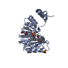
- PDB-6rxr: Crystal structure of CobB Ac2 (A76G, I131C, V162G) in complex wit... -
+ Open data

- Basic information
| Entry | Database: PDB / ID: 6rxr | ||||||
|---|---|---|---|---|---|---|---|
| Title | Crystal structure of CobB Ac2 (A76G, I131C, V162G) in complex with H4K16Cr-2'OH-ADPr peptide intermediate after co-crystallisation | ||||||
 Components Components |
| ||||||
 Keywords Keywords | HYDROLASE / deacylase / NAD-dependent | ||||||
| Function / homology |  Function and homology information Function and homology informationlipoamidase activity / NAD-dependent protein de-2-hydroxyisobutyrylase activity / protein-malonyllysine demalonylase activity / protein-succinyllysine desuccinylase activity / HATs acetylate histones / RNA polymerase I upstream activating factor complex / Condensation of Prophase Chromosomes / : / : / : ...lipoamidase activity / NAD-dependent protein de-2-hydroxyisobutyrylase activity / protein-malonyllysine demalonylase activity / protein-succinyllysine desuccinylase activity / HATs acetylate histones / RNA polymerase I upstream activating factor complex / Condensation of Prophase Chromosomes / : / : / : / Assembly of the ORC complex at the origin of replication / HDACs deacetylate histones / protein acetyllysine N-acetyltransferase / NAD-dependent protein lysine deacetylase activity / cyclic-di-GMP binding / Oxidative Stress Induced Senescence / Recruitment and ATM-mediated phosphorylation of repair and signaling proteins at DNA double strand breaks / RMTs methylate histone arginines / SUMOylation of chromatin organization proteins / RNA Polymerase I Promoter Escape / positive regulation of transcription by RNA polymerase I / nucleolar large rRNA transcription by RNA polymerase I / Estrogen-dependent gene expression / NAD+ poly-ADP-ribosyltransferase activity / NAD+ binding / chemotaxis / structural constituent of chromatin / nucleosome / nucleosome assembly / chromatin organization / defense response to virus / protein heterodimerization activity / regulation of DNA-templated transcription / protein homodimerization activity / DNA binding / zinc ion binding / identical protein binding / nucleus / cytoplasm Similarity search - Function | ||||||
| Biological species |   | ||||||
| Method |  X-RAY DIFFRACTION / SYNCHROTRON / MOLECULAR REPLACEMENT / Resolution: 1.7 Å X-RAY DIFFRACTION / SYNCHROTRON / MOLECULAR REPLACEMENT / Resolution: 1.7 Å | ||||||
 Authors Authors | Spinck, M. / Gasper, R. / Neumann, H. | ||||||
| Funding support |  Germany, 1items Germany, 1items
| ||||||
 Citation Citation | Journal: Angew.Chem.Int.Ed.Engl. / Year: 2020 Title: Evolved, Selective Erasers of Distinct Lysine Acylations. Authors: Spinck, M. / Neumann-Staubitz, P. / Ecke, M. / Gasper, R. / Neumann, H. | ||||||
| History |
|
- Structure visualization
| Structure viewer | Molecule: MolmilJmol/JSmol |
|---|
- Downloads & links
-Download
| PDBx/mmCIF format | 6rxr.cif.gz | 222.1 KB | Display | PDBx/mmCIF format |
|---|---|---|---|---|
| PDB format | pdb6rxr.ent.gz | 176.8 KB | Display | PDB format |
| PDBx/mmJSON format | 6rxr.json.gz | Tree view | PDBx/mmJSON format | |
| Others |  Other downloads Other downloads |
-Validation report
| Arichive directory | https://data.pdbj.org/pub/pdb/validation_reports/rx/6rxrftp://data.pdbj.org/pub/pdb/validation_reports/rx/6rxr | HTTPS FTP |
|---|
-Related structure data
| Related structure data | 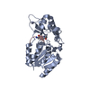 6rxjC 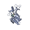 6rxkC 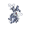 6rxlC 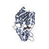 6rxmC  6rxoC 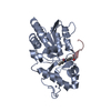 6rxpC  6rxqC 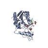 6rxsC  1s5pS S: Starting model for refinement C: citing same article ( |
|---|---|
| Similar structure data |


-Links
 PDBj
PDBj


- Assembly
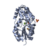
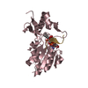
| Deposited unit | 
| |||||||||||||||||||||||||||||||||||||||||||||||||||||||||||||||||||||||||||
|---|---|---|---|---|---|---|---|---|---|---|---|---|---|---|---|---|---|---|---|---|---|---|---|---|---|---|---|---|---|---|---|---|---|---|---|---|---|---|---|---|---|---|---|---|---|---|---|---|---|---|---|---|---|---|---|---|---|---|---|---|---|---|---|---|---|---|---|---|---|---|---|---|---|---|---|---|
| 1 | 
| |||||||||||||||||||||||||||||||||||||||||||||||||||||||||||||||||||||||||||
| 2 | 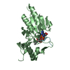
| |||||||||||||||||||||||||||||||||||||||||||||||||||||||||||||||||||||||||||
| 3 | 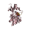
| |||||||||||||||||||||||||||||||||||||||||||||||||||||||||||||||||||||||||||
| 4 | 
| |||||||||||||||||||||||||||||||||||||||||||||||||||||||||||||||||||||||||||
| Unit cell |
| |||||||||||||||||||||||||||||||||||||||||||||||||||||||||||||||||||||||||||
| Noncrystallographic symmetry (NCS) | NCS domain:
NCS domain segments:
NCS ensembles :
|
-Components
| #1: Protein | Mass: 28054.656 Da / Num. of mol.: 4 Source method: isolated from a genetically manipulated source Source: (gene. exp.) Strain: K12 / Gene: cobB, ycfY, b1120, JW1106 / Production host: References: UniProt: P75960, Hydrolases; Acting on carbon-nitrogen bonds, other than peptide bonds; In linear amides #2: Protein/peptide | Mass: 1269.586 Da / Num. of mol.: 4 / Source method: obtained synthetically Source: (synth.) References: UniProt: P02309 #3: Chemical | ChemComp-KMQ / [[( 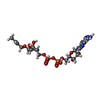  Mass: 613.406 Da / Num. of mol.: 4 / Source method: obtained synthetically / Formula: C19H29N5O14P2 Mass: 613.406 Da / Num. of mol.: 4 / Source method: obtained synthetically / Formula: C19H29N5O14P2#4: Water | ChemComp-HOH / |  Mass: 18.015 Da / Num. of mol.: 582 / Source method: isolated from a natural source / Formula: H2O Mass: 18.015 Da / Num. of mol.: 582 / Source method: isolated from a natural source / Formula: H2OHas ligand of interest | Y | Has protein modification | Y | |
|---|
-Experimental details
-Experiment
| Experiment | Method: X-RAY DIFFRACTION / Number of used crystals: 1 |
|---|
- Sample preparation
| Crystal | Density Matthews: 2.38 Å3/Da / Density % sol: 43.96 % / Description: 48.4 |
|---|---|
| Crystal grow | Temperature: 293 K / Method: vapor diffusion, hanging drop / pH: 7.4 / Details: 0.1 M Bis-Tris, 0.02 M HCl, 22% PEG 3350, 1 mM NAD |
-Data collection
| Diffraction | Mean temperature: 100 K / Serial crystal experiment: N | ||||||||||||||||||||||||||||||||||||||||||||||||||||||||||||||||||||||||||||||||||||||||||||||||||||||||||||||||||||||||||||||||||||||||||||||||||||||||||||||||||||||||||||||||||||||||||||||||||||||||||||||||||
|---|---|---|---|---|---|---|---|---|---|---|---|---|---|---|---|---|---|---|---|---|---|---|---|---|---|---|---|---|---|---|---|---|---|---|---|---|---|---|---|---|---|---|---|---|---|---|---|---|---|---|---|---|---|---|---|---|---|---|---|---|---|---|---|---|---|---|---|---|---|---|---|---|---|---|---|---|---|---|---|---|---|---|---|---|---|---|---|---|---|---|---|---|---|---|---|---|---|---|---|---|---|---|---|---|---|---|---|---|---|---|---|---|---|---|---|---|---|---|---|---|---|---|---|---|---|---|---|---|---|---|---|---|---|---|---|---|---|---|---|---|---|---|---|---|---|---|---|---|---|---|---|---|---|---|---|---|---|---|---|---|---|---|---|---|---|---|---|---|---|---|---|---|---|---|---|---|---|---|---|---|---|---|---|---|---|---|---|---|---|---|---|---|---|---|---|---|---|---|---|---|---|---|---|---|---|---|---|---|---|---|---|
| Diffraction source | Source: SYNCHROTRON / Site: SLS  / Beamline: X10SA / Wavelength: 1 Å / Beamline: X10SA / Wavelength: 1 Å | ||||||||||||||||||||||||||||||||||||||||||||||||||||||||||||||||||||||||||||||||||||||||||||||||||||||||||||||||||||||||||||||||||||||||||||||||||||||||||||||||||||||||||||||||||||||||||||||||||||||||||||||||||
| Detector | Type: DECTRIS PILATUS 6M / Detector: PIXEL / Date: Nov 24, 2018 | ||||||||||||||||||||||||||||||||||||||||||||||||||||||||||||||||||||||||||||||||||||||||||||||||||||||||||||||||||||||||||||||||||||||||||||||||||||||||||||||||||||||||||||||||||||||||||||||||||||||||||||||||||
| Radiation | Protocol: SINGLE WAVELENGTH / Monochromatic (M) / Laue (L): M / Scattering type: x-ray | ||||||||||||||||||||||||||||||||||||||||||||||||||||||||||||||||||||||||||||||||||||||||||||||||||||||||||||||||||||||||||||||||||||||||||||||||||||||||||||||||||||||||||||||||||||||||||||||||||||||||||||||||||
| Radiation wavelength | Wavelength: 1 Å / Relative weight: 1 | ||||||||||||||||||||||||||||||||||||||||||||||||||||||||||||||||||||||||||||||||||||||||||||||||||||||||||||||||||||||||||||||||||||||||||||||||||||||||||||||||||||||||||||||||||||||||||||||||||||||||||||||||||
| Reflection | Resolution: 1.7→45.115 Å / Num. obs: 117240 / % possible obs: 100 % / Redundancy: 6.723 % / Biso Wilson estimate: 32.79 Å2 / CC1/2: 1 / Rmerge(I) obs: 0.047 / Rrim(I) all: 0.051 / Χ2: 1.031 / Net I/σ(I): 20.1 / Num. measured all: 788173 / Scaling rejects: 27 | ||||||||||||||||||||||||||||||||||||||||||||||||||||||||||||||||||||||||||||||||||||||||||||||||||||||||||||||||||||||||||||||||||||||||||||||||||||||||||||||||||||||||||||||||||||||||||||||||||||||||||||||||||
| Reflection shell | Diffraction-ID: 1
|
- Processing
| Software |
| ||||||||||||||||||||||||||||||||||||||||||||||||||||||||||||||||||||||||||||||||||||||||||
|---|---|---|---|---|---|---|---|---|---|---|---|---|---|---|---|---|---|---|---|---|---|---|---|---|---|---|---|---|---|---|---|---|---|---|---|---|---|---|---|---|---|---|---|---|---|---|---|---|---|---|---|---|---|---|---|---|---|---|---|---|---|---|---|---|---|---|---|---|---|---|---|---|---|---|---|---|---|---|---|---|---|---|---|---|---|---|---|---|---|---|---|
| Refinement | Method to determine structure: MOLECULAR REPLACEMENT Starting model: pdbid 1S5P Resolution: 1.7→45.115 Å / SU ML: 0.2 / Cross valid method: THROUGHOUT / σ(F): 1.35 / Phase error: 26.84 / Stereochemistry target values: ML
| ||||||||||||||||||||||||||||||||||||||||||||||||||||||||||||||||||||||||||||||||||||||||||
| Solvent computation | Shrinkage radii: 0.9 Å / VDW probe radii: 1.11 Å / Solvent model: FLAT BULK SOLVENT MODEL | ||||||||||||||||||||||||||||||||||||||||||||||||||||||||||||||||||||||||||||||||||||||||||
| Displacement parameters | Biso max: 118.25 Å2 / Biso mean: 44.7376 Å2 / Biso min: 21.09 Å2 | ||||||||||||||||||||||||||||||||||||||||||||||||||||||||||||||||||||||||||||||||||||||||||
| Refinement step | Cycle: final / Resolution: 1.7→45.115 Å
| ||||||||||||||||||||||||||||||||||||||||||||||||||||||||||||||||||||||||||||||||||||||||||
| Refine LS restraints |
| ||||||||||||||||||||||||||||||||||||||||||||||||||||||||||||||||||||||||||||||||||||||||||
| Refine LS restraints NCS |
| ||||||||||||||||||||||||||||||||||||||||||||||||||||||||||||||||||||||||||||||||||||||||||
| LS refinement shell | Refine-ID: X-RAY DIFFRACTION / Rfactor Rfree error: 0 / Total num. of bins used: 14 / % reflection obs: 100 %
|
