 Movie
Movie Controller

[English] 日本語
 Yorodumi
- PDB-1l7n: TRANSITION STATE ANALOGUE OF PHOSPHOSERINE PHOSPHATASE (ALUMINUM ... -
+ Open data

- Basic information
| Entry | Database: PDB / ID: 1l7n | ||||||
|---|---|---|---|---|---|---|---|
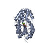
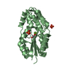
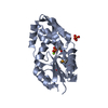
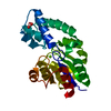
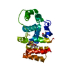
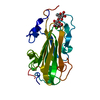
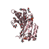
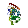
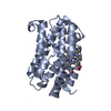
| Title | TRANSITION STATE ANALOGUE OF PHOSPHOSERINE PHOSPHATASE (ALUMINUM FLUORIDE COMPLEX) | ||||||
 Components Components | PHOSPHOSERINE PHOSPHATASE | ||||||
 Keywords Keywords | HYDROLASE / Rossmann fold / b-hairpin / four-Helix bundle / Structural Genomics / BSGC structure funded by NIH / Protein Structure Initiative / PSI / Berkeley Structural Genomics Center | ||||||
| Function / homology |  Function and homology information Function and homology informationphosphoserine phosphatase / L-phosphoserine phosphatase activity / L-serine biosynthetic process / magnesium ion binding / cytoplasm Similarity search - Function | ||||||
| Biological species |   Methanocaldococcus jannaschii (archaea) Methanocaldococcus jannaschii (archaea) | ||||||
| Method |  X-RAY DIFFRACTION / SYNCHROTRON / MOLECULAR REPLACEMENT / Resolution: 1.8 Å X-RAY DIFFRACTION / SYNCHROTRON / MOLECULAR REPLACEMENT / Resolution: 1.8 Å | ||||||
 Authors Authors | Wang, W. / Cho, H.S. / Kim, R. / Jancarik, J. / Yokota, H. / Nguyen, H.H. / Grigoriev, I.V. / Wemmer, D.E. / Kim, S.H. / Berkeley Structural Genomics Center (BSGC) | ||||||
 Citation Citation | Journal: J.Mol.Biol. / Year: 2002 Title: Structural characterization of the reaction pathway in phosphoserine phosphatase: crystallographic "snapshots" of intermediate states. Authors: Wang, W. / Cho, H.S. / Kim, R. / Jancarik, J. / Yokota, H. / Nguyen, H.H. / Grigoriev, I.V. / Wemmer, D.E. / Kim, S.H. | ||||||
| History |
|
- Structure visualization
| Structure viewer | Molecule: MolmilJmol/JSmol |
|---|
- Downloads & links
-Download
| PDBx/mmCIF format | 1l7n.cif.gz | 106.1 KB | Display | PDBx/mmCIF format |
|---|---|---|---|---|
| PDB format | pdb1l7n.ent.gz | 79.8 KB | Display | PDB format |
| PDBx/mmJSON format | 1l7n.json.gz | Tree view | PDBx/mmJSON format | |
| Others |  Other downloads Other downloads |
-Validation report
| Arichive directory | https://data.pdbj.org/pub/pdb/validation_reports/l7/1l7nftp://data.pdbj.org/pub/pdb/validation_reports/l7/1l7n | HTTPS FTP |
|---|
-Related structure data
| Related structure data | 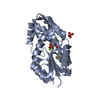 1l7mC 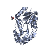 1l7oC 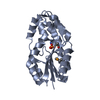 1l7pC 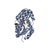 1f5sS S: Starting model for refinement C: citing same article ( |
|---|---|
| Similar structure data | |
| Other databases |


-Links
 PDBj
PDBj


- Assembly
| Deposited unit | 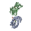
| ||||||||
|---|---|---|---|---|---|---|---|---|---|
| 1 | 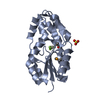
| ||||||||
| 2 | 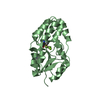
| ||||||||
| Unit cell |
|
-Components
-Protein , 1 types, 2 molecules AB
| #1: Protein | Mass: 23775.346 Da / Num. of mol.: 2 Source method: isolated from a genetically manipulated source Source: (gene. exp.) Methanocaldococcus jannaschii (archaea)Gene: MJ1594 / Plasmid: pET21a / Production host:  |
|---|
-Non-polymers , 5 types, 396 molecules 


| #2: Chemical |  Mass: 102.975 Da / Num. of mol.: 2 / Source method: obtained synthetically / Formula: AlF4 Mass: 102.975 Da / Num. of mol.: 2 / Source method: obtained synthetically / Formula: AlF4#3: Chemical |  Mass: 24.305 Da / Num. of mol.: 2 / Source method: obtained synthetically / Formula: Mg Mass: 24.305 Da / Num. of mol.: 2 / Source method: obtained synthetically / Formula: Mg#4: Chemical | ChemComp-SO4 / |  Mass: 96.063 Da / Num. of mol.: 1 / Source method: obtained synthetically / Formula: SO4 Mass: 96.063 Da / Num. of mol.: 1 / Source method: obtained synthetically / Formula: SO4#5: Chemical |  Mass: 83.977 Da / Num. of mol.: 2 / Source method: obtained synthetically / Formula: AlF3 Mass: 83.977 Da / Num. of mol.: 2 / Source method: obtained synthetically / Formula: AlF3#6: Water | ChemComp-HOH / | Mass: 18.015 Da / Num. of mol.: 389 / Source method: isolated from a natural source / Formula: H2O |
|---|
-Details
| Has protein modification | Y |
|---|
-Experimental details
-Experiment
| Experiment | Method: X-RAY DIFFRACTION / Number of used crystals: 1 |
|---|
- Sample preparation
| Crystal | Density Matthews: 2.68 Å3/Da / Density % sol: 53 % |
|---|---|
| Crystal grow | Temperature: 298 K / Method: vapor diffusion, hanging drop / pH: 4.5 Details: 22% PEG 200 MME, 0.2M SODIUM SULFATE, 0.1M ACETATE BUFFER, 5mM MgCl2, AlCl3, NaF, pH 4.5, VAPOR DIFFUSION, HANGING DROP, temperature 25K |
-Data collection
| Diffraction | Mean temperature: 100 K |
|---|---|
| Diffraction source | Source: SYNCHROTRON / Site: ALS  / Beamline: 5.0.2 / Wavelength: 0.95 Å / Beamline: 5.0.2 / Wavelength: 0.95 Å |
| Detector | Type: ADSC QUANTUM 4 / Detector: CCD / Date: Jun 25, 2001 |
| Radiation | Protocol: SINGLE WAVELENGTH / Monochromatic (M) / Laue (L): M / Scattering type: x-ray |
| Radiation wavelength | Wavelength: 0.95 Å / Relative weight: 1 |
| Reflection | Resolution: 1.8→20 Å / Num. all: 42392 / Num. obs: 41968 / % possible obs: 99 % / Observed criterion σ(I): -3 / Biso Wilson estimate: 20.4 Å2 / Rsym value: 0.06 / Net I/σ(I): 16 |
| Reflection shell | Resolution: 1.8→1.9 Å / Mean I/σ(I) obs: 3.3 / Rsym value: 0.358 / % possible all: 98.7 |
- Processing
| Software |
| |||||||||||||||||||||||||
|---|---|---|---|---|---|---|---|---|---|---|---|---|---|---|---|---|---|---|---|---|---|---|---|---|---|---|
| Refinement | Method to determine structure: MOLECULAR REPLACEMENT Starting model: PDB ENTRY 1F5S Resolution: 1.8→15 Å / Cross valid method: FREE R-VALUE / σ(F): 0 / Stereochemistry target values: CNS
| |||||||||||||||||||||||||
| Displacement parameters |
| |||||||||||||||||||||||||
| Refinement step | Cycle: LAST / Resolution: 1.8→15 Å
|
