Mass: 18.015 Da / Num. of mol.: 274 / Source method: isolated from a natural source / Formula: H2O
Has protein modification
Y
Sequence details
THE CONSTRUCT WAS EXPRESSED WITH A PURIFICATION TAG MGSDKIHHHHHHENLYFQG. THE TAG WAS REMOVED WITH ...THE CONSTRUCT WAS EXPRESSED WITH A PURIFICATION TAG MGSDKIHHHHHHENLYFQG. THE TAG WAS REMOVED WITH TEV PROTEASE LEAVING ONLY A GLYCINE (0) FOLLOWED BY THE TARGET SEQUENCE.
-
Experimental details
-
Experiment
Experiment
Method: X-RAY DIFFRACTION / Number of used crystals: 1
-
Sample preparation
Crystal
Density Matthews: 2.43 Å3/Da / Density % sol: 49.37 %
Crystal grow
Temperature: 277 K / Method: vapor diffusion, sitting drop / pH: 9.5 Details: NANODROP, 1.0M Sodium citrate, 0.1M CHES pH 9.5, VAPOR DIFFUSION, SITTING DROP, temperature 277K
Resolution: 2→29.062 Å / Num. obs: 28773 / % possible obs: 99.9 % / Redundancy: 3.6 % / Biso Wilson estimate: 28.45 Å2 / Rmerge(I) obs: 0.093 / Rsym value: 0.093 / Net I/σ(I): 6.6
Reflection shell
Diffraction-ID: 1
Resolution (Å)
Redundancy (%)
Rmerge(I) obs
Mean I/σ(I) obs
Num. measured all
Num. unique all
Rsym value
% possible all
2-2.05
3.7
0.667
1.1
7762
2095
0.667
100
2.05-2.11
3.6
0.592
1.3
7460
2059
0.592
100
2.11-2.17
3.7
0.479
1.6
7255
1981
0.479
100
2.17-2.24
3.7
0.392
2
7201
1947
0.392
100
2.24-2.31
3.6
0.345
2.2
6766
1862
0.345
100
2.31-2.39
3.7
0.277
2.8
6607
1803
0.277
100
2.39-2.48
3.7
0.229
3.3
6476
1771
0.229
100
2.48-2.58
3.7
0.183
4.2
6265
1706
0.183
100
2.58-2.7
3.7
0.155
4.8
5984
1631
0.155
100
2.7-2.83
3.6
0.123
6.1
5666
1559
0.123
100
2.83-2.98
3.6
0.097
7.5
5448
1493
0.097
100
2.98-3.16
3.7
0.083
8.6
5078
1391
0.083
100
3.16-3.38
3.6
0.069
9.9
4851
1342
0.069
100
3.38-3.65
3.6
0.057
11.6
4517
1254
0.057
99.8
3.65-4
3.6
0.051
12.8
4085
1134
0.051
99.8
4-4.47
3.6
0.046
13.2
3750
1040
0.046
99.6
4.47-5.16
3.6
0.046
12.4
3332
928
0.046
99.5
5.16-6.32
3.5
0.054
11.9
2810
795
0.054
99.4
6.32-8.94
3.5
0.049
12
2166
627
0.049
98.9
8.94-29.062
3.2
0.048
11.7
1136
355
0.048
94.7
-
Phasing
Phasing
Method: MAD
-
Processing
Software
Name
Version
Classification
NB
REFMAC
5.2.0019
refinement
PHENIX
refinement
SHELX
phasing
MolProbity
3beta29
modelbuilding
SCALA
datascaling
PDB_EXTRACT
3
dataextraction
MAR345
CCD
datacollection
MOSFLM
datareduction
autoSHARP
phasing
SHELXD
phasing
Refinement
Method to determine structure: MAD / Resolution: 2→29.062 Å / Cor.coef. Fo:Fc: 0.965 / Cor.coef. Fo:Fc free: 0.947 / SU B: 7.25 / SU ML: 0.105 / TLS residual ADP flag: LIKELY RESIDUAL / Cross valid method: THROUGHOUT / σ(F): 0 / ESU R: 0.164 / ESU R Free: 0.15 Stereochemistry target values: MAXIMUM LIKELIHOOD WITH PHASES Details: 1. HYDROGENS HAVE BEEN ADDED IN THE RIDING POSITIONS. 2. ATOM RECORDS CONTAIN RESIDUAL B FACTORS ONLY. 3. A MET-INHIBITION PROTOCOL WAS USED FOR SELENOMETHIONINE INCORPORATION DURING PROTEIN ...Details: 1. HYDROGENS HAVE BEEN ADDED IN THE RIDING POSITIONS. 2. ATOM RECORDS CONTAIN RESIDUAL B FACTORS ONLY. 3. A MET-INHIBITION PROTOCOL WAS USED FOR SELENOMETHIONINE INCORPORATION DURING PROTEIN EXPRESSION. THE OCCUPANCY OF THE SE ATOMS IN THE MSE RESIDUES WAS REDUCED TO 0.75 FOR THE REDUCED SCATTERING POWER DUE TO PARTIAL S-MET INCORPORATION. 4. 2-[N-CYCLOHEXYLAMINO]ETHANE SULFONIC ACID (NHE) AND CITRATE (CIT) FROM CRYSTALLIZATION AND ETHYLENE GLYCOL (EDO) FROM CRYO SOLUTION WERE MODELED.
Rfactor
Num. reflection
% reflection
Selection details
Rfree
0.212
1455
5.1 %
RANDOM
Rwork
0.167
-
-
-
obs
0.169
28741
99.74 %
-
Solvent computation
Ion probe radii: 0.8 Å / Shrinkage radii: 0.8 Å / VDW probe radii: 1.2 Å / Solvent model: MASK
In the structure databanks used in Yorodumi, some data are registered as the other names, "COVID-19 virus" and "2019-nCoV". Here are the details of the virus and the list of structure data.
Jan 31, 2019. EMDB accession codes are about to change! (news from PDBe EMDB page)
EMDB accession codes are about to change! (news from PDBe EMDB page)
The allocation of 4 digits for EMDB accession codes will soon come to an end. Whilst these codes will remain in use, new EMDB accession codes will include an additional digit and will expand incrementally as the available range of codes is exhausted. The current 4-digit format prefixed with “EMD-” (i.e. EMD-XXXX) will advance to a 5-digit format (i.e. EMD-XXXXX), and so on. It is currently estimated that the 4-digit codes will be depleted around Spring 2019, at which point the 5-digit format will come into force.
The EM Navigator/Yorodumi systems omit the EMD- prefix.
Related info.:Q: What is EMD? / ID/Accession-code notation in Yorodumi/EM Navigator
Yorodumi is a browser for structure data from EMDB, PDB, SASBDB, etc.
This page is also the successor to EM Navigator detail page, and also detail information page/front-end page for Omokage search.
The word "yorodu" (or yorozu) is an old Japanese word meaning "ten thousand". "mi" (miru) is to see.
Related info.:EMDB / PDB / SASBDB / Comparison of 3 databanks / Yorodumi Search / Aug 31, 2016. New EM Navigator & Yorodumi / Yorodumi Papers / Jmol/JSmol / Function and homology information / Changes in new EM Navigator and Yorodumi
 Movie
Movie Controller

 Yorodumi
 Open data
 Basic information
 Components
 Keywords
 Function and homology information
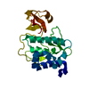
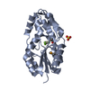
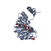
 Corynebacterium glutamicum ATCC 13032 (bacteria)
 X-RAY DIFFRACTION /
  Authors
 Citation
 Structure visualization
 Downloads & links
 Other downloads


 PDBj
PDBj Assembly
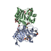


 Mass: 207.290 Da / Num. of mol.: 2 / Source method: obtained synthetically / Formula: C8H17NO3S / Comment: pH buffer*YM
 Mass: 62.068 Da / Num. of mol.: 8 / Source method: obtained synthetically / Formula: C2H6O2
 Mass: 192.124 Da / Num. of mol.: 1 / Source method: obtained synthetically / Formula: C6H8O7
 Mass: 18.015 Da / Num. of mol.: 274 / Source method: isolated from a natural source / Formula: H2O
 Sample preparation
 / Beamline: BL9-2 / Wavelength: 0.91837, 0.97978
 Processing