 Movie
Movie Controller


+ Open data

- Basic information
| Entry | Database: PDB / ID: 2x6q | ||||||
|---|---|---|---|---|---|---|---|
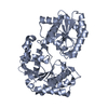
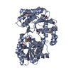
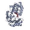
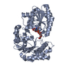
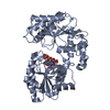
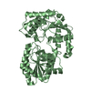
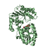
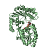
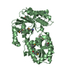
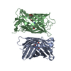
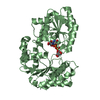
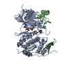
| Title | Crystal structure of trehalose synthase TreT from P.horikoshi | ||||||
 Components Components | TREHALOSE-SYNTHASE TRET | ||||||
 Keywords Keywords | BIOSYNTHETIC PROTEIN | ||||||
| Function / homology |  Function and homology information Function and homology informationalpha,alpha-trehalose synthase / trehalose synthase activity / glucose metabolic process Similarity search - Function | ||||||
| Biological species |   PYROCOCCUS HORIKOSHII (archaea) PYROCOCCUS HORIKOSHII (archaea) | ||||||
| Method |  X-RAY DIFFRACTION / SYNCHROTRON / MOLECULAR REPLACEMENT / Resolution: 2.2 Å X-RAY DIFFRACTION / SYNCHROTRON / MOLECULAR REPLACEMENT / Resolution: 2.2 Å | ||||||
 Authors Authors | Song, H.-N. / Jung, T.-Y. / Yoon, S.-M. / Lim, M.-Y. / Lee, S.-B. / Woo, E.-J. | ||||||
 Citation Citation | Journal: J.Mol.Biol. / Year: 2010 Title: Structural Insights on the New Mechanism of Trehalose Synthesis by Trehalose Synthase Tret from Pyrococcus Horikoshii. Authors: Woo, E.-J. / Ryu, S. / Song, H.-N. / Jung, T.-Y. / Yeon, S. / Lee, H. / Park, B.C. / Park, K. / Lee, S.-B. | ||||||
| History |
|
- Structure visualization
| Structure viewer | Molecule: MolmilJmol/JSmol |
|---|
- Downloads & links
-Download
| PDBx/mmCIF format | 2x6q.cif.gz | 178.1 KB | Display | PDBx/mmCIF format |
|---|---|---|---|---|
| PDB format | pdb2x6q.ent.gz | 143.9 KB | Display | PDB format |
| PDBx/mmJSON format | 2x6q.json.gz | Tree view | PDBx/mmJSON format | |
| Others |  Other downloads Other downloads |
-Validation report
| Arichive directory | https://data.pdbj.org/pub/pdb/validation_reports/x6/2x6qftp://data.pdbj.org/pub/pdb/validation_reports/x6/2x6q | HTTPS FTP |
|---|
-Related structure data


-Links
 PDBj
PDBj
- Assembly
| Deposited unit | 
| ||||||||
|---|---|---|---|---|---|---|---|---|---|
| 1 | 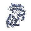
| ||||||||
| 2 | 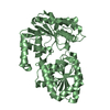
| ||||||||
| Unit cell |
| ||||||||
| Noncrystallographic symmetry (NCS) | NCS oper: (Code: given Matrix: (-0.999803, -0.010977, 0.01653), Vector: |
-Components
| #1: Protein | Mass: 48237.551 Da / Num. of mol.: 2 Source method: isolated from a genetically manipulated source Source: (gene. exp.) PYROCOCCUS HORIKOSHII (archaea) / Plasmid: P6XHIS119 / Production host:  #2: Water | ChemComp-HOH / |  Mass: 18.015 Da / Num. of mol.: 272 / Source method: isolated from a natural source / Formula: H2O Mass: 18.015 Da / Num. of mol.: 272 / Source method: isolated from a natural source / Formula: H2O |
|---|
-Experimental details
-Experiment
| Experiment | Method: X-RAY DIFFRACTION / Number of used crystals: 1 |
|---|
- Sample preparation
| Crystal | Density Matthews: 2.34 Å3/Da / Density % sol: 46.6 % / Description: NONE |
|---|---|
| Crystal grow | Details: PEG 3350 25%, 0.2M MGCL2, 0.1M SODIUM HEPES BUFFER |
-Data collection
| Diffraction | Mean temperature: 297 K |
|---|---|
| Diffraction source | Source: SYNCHROTRON / Site: PAL/PLS  / Beamline: 4A / Wavelength: 1.2399 / Beamline: 4A / Wavelength: 1.2399 |
| Detector | Date: Dec 1, 2005 |
| Radiation | Protocol: SINGLE WAVELENGTH / Monochromatic (M) / Laue (L): M / Scattering type: x-ray |
| Radiation wavelength | Wavelength: 1.2399 Å / Relative weight: 1 |
| Reflection | Resolution: 2.2→29.99 Å / Num. obs: 41687 / % possible obs: 90.5 % / Observed criterion σ(I): 0 / Redundancy: 3.4 % / Biso Wilson estimate: 18.9 Å2 / Rmerge(I) obs: 0.08 / Net I/σ(I): 16.4 |
| Reflection shell | Resolution: 2.2→2.28 Å / Redundancy: 2.6 % / Rmerge(I) obs: 0.34 / Mean I/σ(I) obs: 3.95 / % possible all: 80.9 |
- Processing
| Software |
| ||||||||||||||||||||||||||||||||||||||||||||||||||||||||||||||||||||||||||||||||
|---|---|---|---|---|---|---|---|---|---|---|---|---|---|---|---|---|---|---|---|---|---|---|---|---|---|---|---|---|---|---|---|---|---|---|---|---|---|---|---|---|---|---|---|---|---|---|---|---|---|---|---|---|---|---|---|---|---|---|---|---|---|---|---|---|---|---|---|---|---|---|---|---|---|---|---|---|---|---|---|---|---|
| Refinement | Method to determine structure: MOLECULAR REPLACEMENT / Resolution: 2.2→29.99 Å / Rfactor Rfree error: 0.006 / Data cutoff high absF: 335657.3 / Data cutoff low absF: 0 / Isotropic thermal model: RESTRAINED / Cross valid method: THROUGHOUT / σ(F): 0 / Details: BULK SOLVENT MODEL USED
| ||||||||||||||||||||||||||||||||||||||||||||||||||||||||||||||||||||||||||||||||
| Solvent computation | Solvent model: FLAT MODEL / Bsol: 45.0601 Å2 / ksol: 0.35 e/Å3 | ||||||||||||||||||||||||||||||||||||||||||||||||||||||||||||||||||||||||||||||||
| Displacement parameters | Biso mean: 32.6 Å2
| ||||||||||||||||||||||||||||||||||||||||||||||||||||||||||||||||||||||||||||||||
| Refine analyze |
| ||||||||||||||||||||||||||||||||||||||||||||||||||||||||||||||||||||||||||||||||
| Refinement step | Cycle: LAST / Resolution: 2.2→29.99 Å
| ||||||||||||||||||||||||||||||||||||||||||||||||||||||||||||||||||||||||||||||||
| Refine LS restraints |
| ||||||||||||||||||||||||||||||||||||||||||||||||||||||||||||||||||||||||||||||||
| Refine LS restraints NCS | NCS model details: RESTRAINTS / Rms dev position: 2.2 Å / Weight position: 29.99 | ||||||||||||||||||||||||||||||||||||||||||||||||||||||||||||||||||||||||||||||||
| LS refinement shell | Resolution: 2.2→2.34 Å / Rfactor Rfree error: 0.033 / Total num. of bins used: 6
| ||||||||||||||||||||||||||||||||||||||||||||||||||||||||||||||||||||||||||||||||
| Xplor file |
|
