 Movie
Movie Controller

[English] 日本語
 Yorodumi
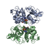
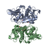
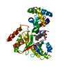
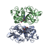
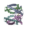
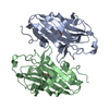
- PDB-3vh6: Crystal structure of the chicken CENP-T histone fold/CENP-W/CENP-... -
+ Open data

- Basic information
| Entry | Database: PDB / ID: 3vh6 | ||||||
|---|---|---|---|---|---|---|---|
| Title | Crystal structure of the chicken CENP-T histone fold/CENP-W/CENP-S/CENP-X heterotetrameric complex, crystal form II | ||||||
 Components Components |
| ||||||
 Keywords Keywords | DNA BINDING PROTEIN / histone fold / chromosome segregation / DNA binding / nucleus | ||||||
| Function / homology |  Function and homology information Function and homology informationPKR-mediated signaling / Amplification of signal from unattached kinetochores via a MAD2 inhibitory signal / RHO GTPases Activate Formins / Resolution of Sister Chromatid Cohesion / EML4 and NUDC in mitotic spindle formation / Deposition of new CENPA-containing nucleosomes at the centromere / Fanconi Anemia Pathway / Separation of Sister Chromatids / FANCM-MHF complex / Fanconi anaemia nuclear complex ...PKR-mediated signaling / Amplification of signal from unattached kinetochores via a MAD2 inhibitory signal / RHO GTPases Activate Formins / Resolution of Sister Chromatid Cohesion / EML4 and NUDC in mitotic spindle formation / Deposition of new CENPA-containing nucleosomes at the centromere / Fanconi Anemia Pathway / Separation of Sister Chromatids / FANCM-MHF complex / Fanconi anaemia nuclear complex / resolution of meiotic recombination intermediates / kinetochore assembly / replication fork processing / chromosome segregation / kinetochore / mitotic cell cycle / protein heterodimerization activity / cell division / DNA repair / chromatin binding / DNA binding / nucleoplasm / nucleus Similarity search - Function | ||||||
| Biological species |  | ||||||
| Method |  X-RAY DIFFRACTION / SYNCHROTRON / MOLECULAR REPLACEMENT / Resolution: 3.351 Å X-RAY DIFFRACTION / SYNCHROTRON / MOLECULAR REPLACEMENT / Resolution: 3.351 Å | ||||||
 Authors Authors | Nishino, T. / Takeuchi, K. / Gascoigne, K.E. / Suzuki, A. / Hori, T. / Oyama, T. / Morikawa, K. / Cheeseman, I.M. / Fukagawa, T. | ||||||
 Citation Citation | Journal: Cell(Cambridge,Mass.) / Year: 2012 Title: CENP-T-W-S-X Forms a Unique Centromeric Chromatin Structure with a Histone-like Fold Authors: Nishino, T. / Takeuchi, K. / Gascoigne, K.E. / Suzuki, A. / Hori, T. / Oyama, T. / Morikawa, K. / Cheeseman, I.M. / Fukagawa, T. | ||||||
| History |
|
- Structure visualization
| Structure viewer | Molecule: MolmilJmol/JSmol |
|---|
- Downloads & links
-Download
| PDBx/mmCIF format | 3vh6.cif.gz | 81.5 KB | Display | PDBx/mmCIF format |
|---|---|---|---|---|
| PDB format | pdb3vh6.ent.gz | 61.9 KB | Display | PDB format |
| PDBx/mmJSON format | 3vh6.json.gz | Tree view | PDBx/mmJSON format | |
| Others |  Other downloads Other downloads |
-Validation report
| Arichive directory | https://data.pdbj.org/pub/pdb/validation_reports/vh/3vh6ftp://data.pdbj.org/pub/pdb/validation_reports/vh/3vh6 | HTTPS FTP |
|---|
-Related structure data
| Related structure data | 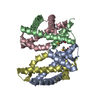 3b0bC 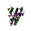 3b0cC 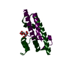 3b0dC 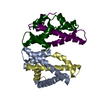 3vh5SC S: Starting model for refinement C: citing same article ( |
|---|---|
| Similar structure data |


-Links
 PDBj
PDBj

- Assembly
| Deposited unit | 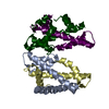
| ||||||||
|---|---|---|---|---|---|---|---|---|---|
| 1 |
| ||||||||
| Unit cell |
|
-Components
| #1: Protein | Mass: 15561.421 Da / Num. of mol.: 1 / Mutation: C26A, C28A, C55A Source method: isolated from a genetically manipulated source Source: (gene. exp.)  |
|---|---|
| #2: Protein | Mass: 9361.746 Da / Num. of mol.: 1 Source method: isolated from a genetically manipulated source Source: (gene. exp.) |
| #3: Protein | Mass: 12682.790 Da / Num. of mol.: 1 / Fragment: C-terminal histone fold / Mutation: C87A, C161A Source method: isolated from a genetically manipulated source Source: (gene. exp.) |
| #4: Protein | Mass: 8855.593 Da / Num. of mol.: 1 Source method: isolated from a genetically manipulated source Source: (gene. exp.) |
| Sequence details | THE SEQUENCE DATABASE REFERENCES |
-Experimental details
-Experiment
| Experiment | Method: X-RAY DIFFRACTION / Number of used crystals: 1 |
|---|
- Sample preparation
| Crystal | Density Matthews: 3.57 Å3/Da / Density % sol: 65.56 % |
|---|---|
| Crystal grow | Temperature: 293 K / Method: vapor diffusion, sitting drop / pH: 8.5 Details: 0.1M Tris-HCl, 5.6% PEG 8000, pH 8.5, VAPOR DIFFUSION, SITTING DROP, temperature 293K |
-Data collection
| Diffraction | Mean temperature: 100 K |
|---|---|
| Diffraction source | Source: SYNCHROTRON / Site: SPring-8  / Beamline: BL44XU / Wavelength: 0.9 Å / Beamline: BL44XU / Wavelength: 0.9 Å |
| Detector | Type: ADSC QUANTUM 210 / Detector: CCD / Date: Jun 15, 2011 |
| Radiation | Monochromator: double-crystal monochromator / Protocol: SINGLE WAVELENGTH / Monochromatic (M) / Laue (L): M / Scattering type: x-ray |
| Radiation wavelength | Wavelength: 0.9 Å / Relative weight: 1 |
| Reflection | Resolution: 3.35→37.4 Å / Num. all: 9985 / Num. obs: 9626 / % possible obs: 99.4 % / Observed criterion σ(F): 1 / Observed criterion σ(I): 1 / Redundancy: 7.3 % / Biso Wilson estimate: 115.15 Å2 |
| Reflection shell | Resolution: 3.35→3.47 Å / Redundancy: 7 % / Rmerge(I) obs: 0.68 / Mean I/σ(I) obs: 2.88 / Num. unique all: 938 / Rsym value: 0.726 / % possible all: 100 |
- Processing
| Software |
| |||||||||||||||||||||||||||||||||||||||||||||||||||||||||||||||||||||||||||||||||||||||||||||||||||||||||
|---|---|---|---|---|---|---|---|---|---|---|---|---|---|---|---|---|---|---|---|---|---|---|---|---|---|---|---|---|---|---|---|---|---|---|---|---|---|---|---|---|---|---|---|---|---|---|---|---|---|---|---|---|---|---|---|---|---|---|---|---|---|---|---|---|---|---|---|---|---|---|---|---|---|---|---|---|---|---|---|---|---|---|---|---|---|---|---|---|---|---|---|---|---|---|---|---|---|---|---|---|---|---|---|---|---|---|
| Refinement | Method to determine structure: MOLECULAR REPLACEMENT Starting model: 3VH5 Resolution: 3.351→37.361 Å / Occupancy max: 1 / Occupancy min: 1 / FOM work R set: 0.7888 / SU ML: 1.09 / σ(F): 1.34 / Phase error: 27.69 / Stereochemistry target values: ML
| |||||||||||||||||||||||||||||||||||||||||||||||||||||||||||||||||||||||||||||||||||||||||||||||||||||||||
| Solvent computation | Shrinkage radii: 0.83 Å / VDW probe radii: 1.1 Å / Solvent model: FLAT BULK SOLVENT MODEL / Bsol: 95.058 Å2 / ksol: 0.326 e/Å3 | |||||||||||||||||||||||||||||||||||||||||||||||||||||||||||||||||||||||||||||||||||||||||||||||||||||||||
| Displacement parameters | Biso max: 352.23 Å2 / Biso mean: 117.7597 Å2 / Biso min: 59.68 Å2
| |||||||||||||||||||||||||||||||||||||||||||||||||||||||||||||||||||||||||||||||||||||||||||||||||||||||||
| Refinement step | Cycle: LAST / Resolution: 3.351→37.361 Å
| |||||||||||||||||||||||||||||||||||||||||||||||||||||||||||||||||||||||||||||||||||||||||||||||||||||||||
| Refine LS restraints |
| |||||||||||||||||||||||||||||||||||||||||||||||||||||||||||||||||||||||||||||||||||||||||||||||||||||||||
| LS refinement shell | Refine-ID: X-RAY DIFFRACTION / Total num. of bins used: 14
|
