 Movie
Movie Controller


+ Open data

- Basic information
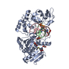
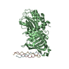
| Entry | Database: PDB / ID: 7bee | ||||||
|---|---|---|---|---|---|---|---|
| Title | Crystal structure of a Hsp47-collagen peptide complex | ||||||
 Components Components |
| ||||||
 Keywords Keywords | CHAPERONE / hsp / hsp47 / collagen | ||||||
| Function / homology |  Function and homology information Function and homology informationCollagen biosynthesis and modifying enzymes / negative regulation of endopeptidase activity / collagen fibril organization / collagen binding / serine-type endopeptidase inhibitor activity / endoplasmic reticulum / extracellular space Similarity search - Function | ||||||
| Biological species |   Homo sapiens (human) Homo sapiens (human) | ||||||
| Method |  X-RAY DIFFRACTION / SYNCHROTRON / MOLECULAR REPLACEMENT / molecular replacement / Resolution: 1.939 Å X-RAY DIFFRACTION / SYNCHROTRON / MOLECULAR REPLACEMENT / molecular replacement / Resolution: 1.939 Å | ||||||
 Authors Authors | Abraham, E.T. / Gebauer, J.M. / Baumann, U. | ||||||
| Funding support |  Germany, 1items Germany, 1items
| ||||||
 Citation Citation | Journal: J.Biol.Chem. / Year: 2021 Title: Collagen's primary structure determines collagen:HSP47 complex stoichiometry. Authors: Abraham, E.T. / Oecal, S. / Morgelin, M. / Schmid, P.W.N. / Buchner, J. / Baumann, U. / Gebauer, J.M. | ||||||
| History |
|
- Structure visualization
| Structure viewer | Molecule: MolmilJmol/JSmol |
|---|
- Downloads & links
-Download
| PDBx/mmCIF format | 7bee.cif.gz | 353.3 KB | Display | PDBx/mmCIF format |
|---|---|---|---|---|
| PDB format | pdb7bee.ent.gz | 291.3 KB | Display | PDB format |
| PDBx/mmJSON format | 7bee.json.gz | Tree view | PDBx/mmJSON format | |
| Others |  Other downloads Other downloads |
-Validation report
| Summary document | 7bee_validation.pdf.gz | 482.1 KB | Display | wwPDB validaton report |
|---|---|---|---|---|
| Full document | 7bee_full_validation.pdf.gz | 505 KB | Display | |
| Data in XML | 7bee_validation.xml.gz | 35.8 KB | Display | |
| Data in CIF | 7bee_validation.cif.gz | 50.9 KB | Display | |
| Arichive directory | https://data.pdbj.org/pub/pdb/validation_reports/be/7beeftp://data.pdbj.org/pub/pdb/validation_reports/be/7bee | HTTPS FTP |
-Related structure data
| Related structure data |  7bduC  7bfiC  4au2S S: Starting model for refinement C: citing same article ( |
|---|---|
| Similar structure data |


-Links
 PDBj
PDBj


- Assembly
| Deposited unit | 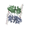
| |||||||||||||||||||||||||||||||||||||||||||||||||||||||||||||||||||||||||||||||||||||||||||||||||||||||||||||||||||||||
|---|---|---|---|---|---|---|---|---|---|---|---|---|---|---|---|---|---|---|---|---|---|---|---|---|---|---|---|---|---|---|---|---|---|---|---|---|---|---|---|---|---|---|---|---|---|---|---|---|---|---|---|---|---|---|---|---|---|---|---|---|---|---|---|---|---|---|---|---|---|---|---|---|---|---|---|---|---|---|---|---|---|---|---|---|---|---|---|---|---|---|---|---|---|---|---|---|---|---|---|---|---|---|---|---|---|---|---|---|---|---|---|---|---|---|---|---|---|---|---|---|
| 1 | 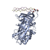
| |||||||||||||||||||||||||||||||||||||||||||||||||||||||||||||||||||||||||||||||||||||||||||||||||||||||||||||||||||||||
| 2 |
| |||||||||||||||||||||||||||||||||||||||||||||||||||||||||||||||||||||||||||||||||||||||||||||||||||||||||||||||||||||||
| 3 | 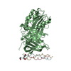
| |||||||||||||||||||||||||||||||||||||||||||||||||||||||||||||||||||||||||||||||||||||||||||||||||||||||||||||||||||||||
| Unit cell |
| |||||||||||||||||||||||||||||||||||||||||||||||||||||||||||||||||||||||||||||||||||||||||||||||||||||||||||||||||||||||
| Noncrystallographic symmetry (NCS) | NCS domain:
NCS domain segments:
|