 Movie
Movie Controller

[English] 日本語
 Yorodumi
- PDB-2np7: Crystal structure of the adenine-specific DNA methyltransferase M... -
+ Open data

- Basic information
| Entry | Database: PDB / ID: 2np7 | ||||||
|---|---|---|---|---|---|---|---|
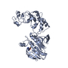
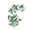
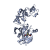
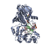
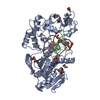
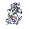
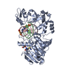
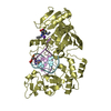
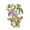
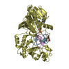
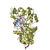
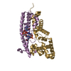
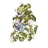
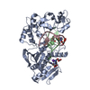
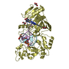
| Title | Crystal structure of the adenine-specific DNA methyltransferase M.TaqI complexed with the cofactor analog AETA and a 10 bp DNA containing an abasic site analog at the target position and pyrrolo-dC at the target base partner position | ||||||
 Components Components |
| ||||||
 Keywords Keywords | TRANSFERASE/DNA / DNA / DNA methyltransferase / target base partner / pyrrolo-dC / abasic site analog / base flipping / nucleotide flipping / TRANSFERASE-DNA COMPLEX | ||||||
| Function / homology |  Function and homology information Function and homology informationsite-specific DNA-methyltransferase (adenine-specific) / site-specific DNA-methyltransferase (adenine-specific) activity / DNA restriction-modification system / methylation / DNA binding Similarity search - Function | ||||||
| Biological species |   Thermus aquaticus (bacteria) Thermus aquaticus (bacteria) | ||||||
| Method |  X-RAY DIFFRACTION / SYNCHROTRON / MOLECULAR REPLACEMENT / Resolution: 1.9 Å X-RAY DIFFRACTION / SYNCHROTRON / MOLECULAR REPLACEMENT / Resolution: 1.9 Å | ||||||
 Authors Authors | Lenz, T. / Scheidig, A.J. / Weinhold, E. | ||||||
 Citation Citation | Journal: To be Published Title: Caught in the act II: Visualization of an intermediate in the DNA base-flipping pathway induced by the adenine-specific DNA methyltransferase M.TaqI Authors: Lenz, T. / Scheidig, A.J. / Weinhold, E. #1: Journal: NAT.STRUCT.BIOL. / Year: 2001Title: Structure of the N6-adenine DNA methyltransferase M.Taqi in complex with DNA and a cofactor analog Authors: Goedecke, K. / Pignot, M. / Goody, R.S. / Scheidig, A.J. / Weinhold, E. #2: Journal: J.Mol.Biol. / Year: 1997Title: Differential binding of s-adenosylmethionine, s-adenosylhomocysteine and sinefungin to the adenine-specific DNA methyltransferase M.Taqi Authors: Schluckebier, G. / Kozak, M. / Bleimling, N. / Weinhold, E. / Saenger, W. | ||||||
| History |
|
- Structure visualization
| Structure viewer | Molecule: MolmilJmol/JSmol |
|---|
- Downloads & links
-Download
| PDBx/mmCIF format | 2np7.cif.gz | 122.5 KB | Display | PDBx/mmCIF format |
|---|---|---|---|---|
| PDB format | pdb2np7.ent.gz | 88.5 KB | Display | PDB format |
| PDBx/mmJSON format | 2np7.json.gz | Tree view | PDBx/mmJSON format | |
| Others |  Other downloads Other downloads |
-Validation report
| Arichive directory | https://data.pdbj.org/pub/pdb/validation_reports/np/2np7ftp://data.pdbj.org/pub/pdb/validation_reports/np/2np7 | HTTPS FTP |
|---|
-Related structure data
| Related structure data | 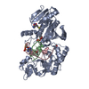 2np6C  1g38S S: Starting model for refinement C: citing same article ( |
|---|---|
| Similar structure data |


-Links
 PDBj
PDBj


- Assembly
| Deposited unit | 
| ||||||||
|---|---|---|---|---|---|---|---|---|---|
| 1 |
| ||||||||
| Unit cell |
| ||||||||
| Details | The asymmetric unit contains one independent biological assembly consisting of M.TaqI, DNA and cofactor analog AETA. |
-Components
-DNA chain , 2 types, 2 molecules BC
| #1: DNA chain | Mass: 2917.890 Da / Num. of mol.: 1 / Source method: obtained synthetically / Details: Solid-phase DNA synthesis |
|---|---|
| #2: DNA chain | Mass: 3075.083 Da / Num. of mol.: 1 / Source method: obtained synthetically / Details: Solid-phase DNA synthesis |
-Protein , 1 types, 1 molecules A
| #3: Protein | Mass: 47931.195 Da / Num. of mol.: 1 Source method: isolated from a genetically manipulated source Source: (gene. exp.) Thermus aquaticus (bacteria) / Strain: YT1 / Gene: taqIM / Plasmid: PA1/MTAQ-A49A / Production host: References: UniProt: P14385, site-specific DNA-methyltransferase (adenine-specific) |
|---|
-Non-polymers , 3 types, 530 molecules 


| #4: Chemical | ChemComp-NEA /  Mass: 326.375 Da / Num. of mol.: 1 / Source method: obtained synthetically / Formula: C12H18N6O3S Mass: 326.375 Da / Num. of mol.: 1 / Source method: obtained synthetically / Formula: C12H18N6O3S | ||
|---|---|---|---|
| #5: Chemical |  Mass: 92.094 Da / Num. of mol.: 2 / Source method: obtained synthetically / Formula: C3H8O3 Mass: 92.094 Da / Num. of mol.: 2 / Source method: obtained synthetically / Formula: C3H8O3#6: Water | ChemComp-HOH / | Mass: 18.015 Da / Num. of mol.: 527 / Source method: isolated from a natural source / Formula: H2O |
-Experimental details
-Experiment
| Experiment | Method: X-RAY DIFFRACTION / Number of used crystals: 1 |
|---|
- Sample preparation
| Crystal | Density Matthews: 2.23 Å3/Da / Density % sol: 44.87 % | ||||||||||||||||||||||||||||||||||||
|---|---|---|---|---|---|---|---|---|---|---|---|---|---|---|---|---|---|---|---|---|---|---|---|---|---|---|---|---|---|---|---|---|---|---|---|---|---|
| Crystal grow | Temperature: 298 K / Method: vapor diffusion, hanging drop / pH: 7.3 Details: 3 microliters crystallization buffer (10 mM Tris/HCl, 300 mM NaCl, pH 7.3) containing the complex plus 1 microliter reservoir solution (100 mM KCl, 100 mM MgCl2, 6% isopropanol, 50 mM sodium ...Details: 3 microliters crystallization buffer (10 mM Tris/HCl, 300 mM NaCl, pH 7.3) containing the complex plus 1 microliter reservoir solution (100 mM KCl, 100 mM MgCl2, 6% isopropanol, 50 mM sodium cacodylate, pH 6.0), VAPOR DIFFUSION, HANGING DROP, temperature 298K | ||||||||||||||||||||||||||||||||||||
| Components of the solutions |
|
-Data collection
| Diffraction | Mean temperature: 100 K |
|---|---|
| Diffraction source | Source: SYNCHROTRON / Site: ESRF  / Beamline: ID14-3 / Wavelength: 0.931 / Beamline: ID14-3 / Wavelength: 0.931 |
| Detector | Type: ADSC QUANTUM 4 / Detector: CCD / Date: May 13, 2005 / Details: ID14-3 (mirror) |
| Radiation | Monochromator: ID14-3 (mirror) / Protocol: SINGLE WAVELENGTH / Monochromatic (M) / Laue (L): M / Scattering type: x-ray |
| Radiation wavelength | Wavelength: 0.931 Å / Relative weight: 1 |
| Reflection | Resolution: 1.9→19.93 Å / Num. all: 39135 / Num. obs: 39083 / % possible obs: 99.9 % / Observed criterion σ(F): 0 / Observed criterion σ(I): 0 / Redundancy: 7.29 % / Biso Wilson estimate: 23.56 Å2 / Rmerge(I) obs: 0.194 / Rsym value: 0.194 / Net I/σ(I): 7.88 |
| Reflection shell | Resolution: 1.9→2 Å / Redundancy: 7.33 % / Rmerge(I) obs: 0.747 / Mean I/σ(I) obs: 2.77 / Num. unique all: 5504 / Rsym value: 0.747 / % possible all: 100 |
- Processing
| Software |
| ||||||||||||||||||||||||||||||||||||||||||||||||||||||||||||||||||||||||||||||||||||||||||||||||||||||||||||||||||||||||||||||||||||||||||||||||||||||||||||||||||||||||||
|---|---|---|---|---|---|---|---|---|---|---|---|---|---|---|---|---|---|---|---|---|---|---|---|---|---|---|---|---|---|---|---|---|---|---|---|---|---|---|---|---|---|---|---|---|---|---|---|---|---|---|---|---|---|---|---|---|---|---|---|---|---|---|---|---|---|---|---|---|---|---|---|---|---|---|---|---|---|---|---|---|---|---|---|---|---|---|---|---|---|---|---|---|---|---|---|---|---|---|---|---|---|---|---|---|---|---|---|---|---|---|---|---|---|---|---|---|---|---|---|---|---|---|---|---|---|---|---|---|---|---|---|---|---|---|---|---|---|---|---|---|---|---|---|---|---|---|---|---|---|---|---|---|---|---|---|---|---|---|---|---|---|---|---|---|---|---|---|---|---|---|---|
| Refinement | Method to determine structure: MOLECULAR REPLACEMENT Starting model: PDB ENTRY 1G38 Resolution: 1.9→19.93 Å / Cor.coef. Fo:Fc: 0.953 / Cor.coef. Fo:Fc free: 0.928 / SU B: 3.331 / SU ML: 0.099 / Cross valid method: THROUGHOUT / σ(F): 0 / ESU R: 0.154 / ESU R Free: 0.142 / Stereochemistry target values: MAXIMUM LIKELIHOOD / Details: HYDROGENS HAVE BEEN ADDED IN THE RIDING POSITIONS
| ||||||||||||||||||||||||||||||||||||||||||||||||||||||||||||||||||||||||||||||||||||||||||||||||||||||||||||||||||||||||||||||||||||||||||||||||||||||||||||||||||||||||||
| Solvent computation | Ion probe radii: 0.8 Å / Shrinkage radii: 0.8 Å / VDW probe radii: 1.2 Å / Solvent model: MASK | ||||||||||||||||||||||||||||||||||||||||||||||||||||||||||||||||||||||||||||||||||||||||||||||||||||||||||||||||||||||||||||||||||||||||||||||||||||||||||||||||||||||||||
| Displacement parameters | Biso mean: 19.777 Å2
| ||||||||||||||||||||||||||||||||||||||||||||||||||||||||||||||||||||||||||||||||||||||||||||||||||||||||||||||||||||||||||||||||||||||||||||||||||||||||||||||||||||||||||
| Refinement step | Cycle: LAST / Resolution: 1.9→19.93 Å
| ||||||||||||||||||||||||||||||||||||||||||||||||||||||||||||||||||||||||||||||||||||||||||||||||||||||||||||||||||||||||||||||||||||||||||||||||||||||||||||||||||||||||||
| Refine LS restraints |
| ||||||||||||||||||||||||||||||||||||||||||||||||||||||||||||||||||||||||||||||||||||||||||||||||||||||||||||||||||||||||||||||||||||||||||||||||||||||||||||||||||||||||||
| LS refinement shell | Resolution: 1.9→1.949 Å / Total num. of bins used: 20
|
