 Movie
Movie Controller

[English] 日本語
 Yorodumi
- PDB-4hym: Pyrrolopyrimidine inhibitors of dna gyrase b and topoisomerase iv... -
+ Open data

- Basic information
| Entry | Database: PDB / ID: 4hym | ||||||
|---|---|---|---|---|---|---|---|
| Title | Pyrrolopyrimidine inhibitors of dna gyrase b and topoisomerase iv, part i: structure guided discovery and optimization of dual targeting agents with potent, broad-spectrum enzymatic activity. | ||||||
 Components Components | Topoisomerase IV, subunit B | ||||||
 Keywords Keywords | Isomerase/Isomerase inhibitor / ATP-binding / Nucleotide-binding / Topoisomerase / ATP-binding domain / Isomerase-Isomerase inhibitor complex | ||||||
| Function / homology |  Function and homology information Function and homology informationDNA topoisomerase type II (double strand cut, ATP-hydrolyzing) activity / DNA topoisomerase (ATP-hydrolysing) / DNA topological change / chromosome segregation / chromosome / DNA binding / ATP binding / metal ion binding Similarity search - Function | ||||||
| Biological species |  Francisella tularensis subsp. holarctica LVS (bacteria) Francisella tularensis subsp. holarctica LVS (bacteria) | ||||||
| Method |  X-RAY DIFFRACTION / SYNCHROTRON / MOLECULAR REPLACEMENT / Resolution: 1.9 Å X-RAY DIFFRACTION / SYNCHROTRON / MOLECULAR REPLACEMENT / Resolution: 1.9 Å | ||||||
 Authors Authors | Bensen, D.C. / Creighton, C.J. / Kwan, B. / Tari, L.W. | ||||||
 Citation Citation | Journal: Bioorg.Med.Chem.Lett. / Year: 2013 Title: Pyrrolopyrimidine inhibitors of DNA gyrase B (GyrB) and topoisomerase IV (ParE). Part I: Structure guided discovery and optimization of dual targeting agents with potent, broad-spectrum enzymatic activity. Authors: Tari, L.W. / Trzoss, M. / Bensen, D.C. / Li, X. / Chen, Z. / Lam, T. / Zhang, J. / Creighton, C.J. / Cunningham, M.L. / Kwan, B. / Stidham, M. / Shaw, K.J. / Lightstone, F.C. / Wong, S.E. / ...Authors: Tari, L.W. / Trzoss, M. / Bensen, D.C. / Li, X. / Chen, Z. / Lam, T. / Zhang, J. / Creighton, C.J. / Cunningham, M.L. / Kwan, B. / Stidham, M. / Shaw, K.J. / Lightstone, F.C. / Wong, S.E. / Nguyen, T.B. / Nix, J. / Finn, J. | ||||||
| History |
|
- Structure visualization
| Structure viewer | Molecule: MolmilJmol/JSmol |
|---|
- Downloads & links
-Download
| PDBx/mmCIF format | 4hym.cif.gz | 146.3 KB | Display | PDBx/mmCIF format |
|---|---|---|---|---|
| PDB format | pdb4hym.ent.gz | 111.6 KB | Display | PDB format |
| PDBx/mmJSON format | 4hym.json.gz | Tree view | PDBx/mmJSON format | |
| Others |  Other downloads Other downloads |
-Validation report
| Arichive directory | https://data.pdbj.org/pub/pdb/validation_reports/hy/4hymftp://data.pdbj.org/pub/pdb/validation_reports/hy/4hym | HTTPS FTP |
|---|
-Related structure data
| Related structure data |  4geeC  4gfnC  4gglC  4hxwC  4hxzC  4hy1C  4hypC  4hz0C  4hz5C C: citing same article ( |
|---|---|
| Similar structure data |


-Links
 PDBj
PDBj


- Assembly
| Deposited unit | 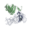
| ||||||||
|---|---|---|---|---|---|---|---|---|---|
| 1 | 
| ||||||||
| 2 | 
| ||||||||
| Unit cell |
|
-Components
| #1: Protein | Mass: 43950.766 Da / Num. of mol.: 2 Source method: isolated from a genetically manipulated source Source: (gene. exp.) Francisella tularensis subsp. holarctica LVS (bacteria)Strain: LVS / Gene: FTL_1726 / Production host: References: UniProt: Q2A1P5, UniProt: A0A0J9WZF0*PLUS, EC: 5.99.1.3 #2: Chemical | ChemComp-CJC / |   Mass: 442.925 Da / Num. of mol.: 1 / Source method: obtained synthetically / Formula: C19H19ClN8OS Mass: 442.925 Da / Num. of mol.: 1 / Source method: obtained synthetically / Formula: C19H19ClN8OS#3: Water | ChemComp-HOH / |  Mass: 18.015 Da / Num. of mol.: 634 / Source method: isolated from a natural source / Formula: H2O Mass: 18.015 Da / Num. of mol.: 634 / Source method: isolated from a natural source / Formula: H2OSequence details | THE RESIDUE AT THIS POSITION INDICATES A NATURAL VARIANT | |
|---|
-Experimental details
-Experiment
| Experiment | Method: X-RAY DIFFRACTION / Number of used crystals: 1 |
|---|
- Sample preparation
| Crystal | Density Matthews: 2.6 Å3/Da / Density % sol: 52.71 % |
|---|---|
| Crystal grow | Temperature: 277 K / pH: 5.4 Details: 10% PEG 4000, 10% isopropanol, 100 mM citrate , pH 5.4, VAPOR DIFFUSION, HANGING DROP, temperature 277K |
-Data collection
| Diffraction | Mean temperature: 100 K |
|---|---|
| Diffraction source | Source: SYNCHROTRON / Site: ALS  / Beamline: 4.2.2 / Wavelength: 1 / Beamline: 4.2.2 / Wavelength: 1 |
| Detector | Type: NOIR-1 / Detector: CCD / Date: Jul 13, 2009 |
| Radiation | Protocol: SINGLE WAVELENGTH / Monochromatic (M) / Laue (L): M / Scattering type: x-ray |
| Radiation wavelength | Wavelength: 1 Å / Relative weight: 1 |
| Reflection | Resolution: 1.9→43.43 Å / Num. obs: 70176 / % possible obs: 99.4 % / Rmerge(I) obs: 0.082 / Net I/σ(I): 7.2 |
| Reflection shell | Resolution: 1.9→1.97 Å / Rmerge(I) obs: 0.414 / Mean I/σ(I) obs: 2.1 / % possible all: 99.1 |
- Processing
| Software |
| ||||||||||||||||||||||||||||||||||||||||||||||||||||||||||||||||||||||||||||||||||||||||||||||||||||||||||||||||||||||||||||||||||||||||||||||||||||||||||||||||||||||||||
|---|---|---|---|---|---|---|---|---|---|---|---|---|---|---|---|---|---|---|---|---|---|---|---|---|---|---|---|---|---|---|---|---|---|---|---|---|---|---|---|---|---|---|---|---|---|---|---|---|---|---|---|---|---|---|---|---|---|---|---|---|---|---|---|---|---|---|---|---|---|---|---|---|---|---|---|---|---|---|---|---|---|---|---|---|---|---|---|---|---|---|---|---|---|---|---|---|---|---|---|---|---|---|---|---|---|---|---|---|---|---|---|---|---|---|---|---|---|---|---|---|---|---|---|---|---|---|---|---|---|---|---|---|---|---|---|---|---|---|---|---|---|---|---|---|---|---|---|---|---|---|---|---|---|---|---|---|---|---|---|---|---|---|---|---|---|---|---|---|---|---|---|
| Refinement | Method to determine structure: MOLECULAR REPLACEMENT / Resolution: 1.9→43.43 Å / Cor.coef. Fo:Fc: 0.936 / Cor.coef. Fo:Fc free: 0.918 / SU B: 3.636 / SU ML: 0.108 / Cross valid method: THROUGHOUT / ESU R: 0.158 / ESU R Free: 0.151 / Stereochemistry target values: MAXIMUM LIKELIHOOD
| ||||||||||||||||||||||||||||||||||||||||||||||||||||||||||||||||||||||||||||||||||||||||||||||||||||||||||||||||||||||||||||||||||||||||||||||||||||||||||||||||||||||||||
| Solvent computation | Ion probe radii: 0.8 Å / Shrinkage radii: 0.8 Å / VDW probe radii: 1.2 Å / Solvent model: BABINET MODEL WITH MASK | ||||||||||||||||||||||||||||||||||||||||||||||||||||||||||||||||||||||||||||||||||||||||||||||||||||||||||||||||||||||||||||||||||||||||||||||||||||||||||||||||||||||||||
| Displacement parameters | Biso mean: 37.92 Å2
| ||||||||||||||||||||||||||||||||||||||||||||||||||||||||||||||||||||||||||||||||||||||||||||||||||||||||||||||||||||||||||||||||||||||||||||||||||||||||||||||||||||||||||
| Refinement step | Cycle: LAST / Resolution: 1.9→43.43 Å
| ||||||||||||||||||||||||||||||||||||||||||||||||||||||||||||||||||||||||||||||||||||||||||||||||||||||||||||||||||||||||||||||||||||||||||||||||||||||||||||||||||||||||||
| Refine LS restraints |
| ||||||||||||||||||||||||||||||||||||||||||||||||||||||||||||||||||||||||||||||||||||||||||||||||||||||||||||||||||||||||||||||||||||||||||||||||||||||||||||||||||||||||||
| LS refinement shell | Resolution: 1.9→1.95 Å / Total num. of bins used: 20
|
