 Movie
Movie Controller


+ Open data

- Basic information
| Entry | Database: PDB / ID: 3doh | ||||||
|---|---|---|---|---|---|---|---|
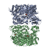
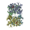
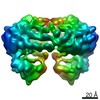
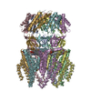
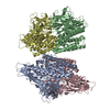
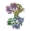
| Title | Crystal Structure of a Thermostable Esterase | ||||||
 Components Components | esterase | ||||||
 Keywords Keywords | HYDROLASE / alpha-beta Hydrolase / beta sheet | ||||||
| Function / homology |  Function and homology information Function and homology information | ||||||
| Biological species |   Thermotoga maritima (bacteria) Thermotoga maritima (bacteria) | ||||||
| Method |  X-RAY DIFFRACTION / SYNCHROTRON / MAD / Resolution: 2.6 Å X-RAY DIFFRACTION / SYNCHROTRON / MAD / Resolution: 2.6 Å | ||||||
 Authors Authors | Levisson, M. / Sun, L. / Hendriks, S. / Dijkstra, B.W. / Van der Oost, J. / Kengen, S.W.M. | ||||||
 Citation Citation | Journal: J.Mol.Biol. / Year: 2009 Title: Crystal structure and biochemical properties of a novel thermostable esterase containing an immunoglobulin-like domain. Authors: Levisson, M. / Sun, L. / Hendriks, S. / Swinkels, P. / Akveld, T. / Bultema, J.B. / Barendregt, A. / van den Heuvel, R.H.H. / Dijkstra, B.W. / van der Oost, J. / Kengen, S.W.M. | ||||||
| History |
|
- Structure visualization
| Structure viewer | Molecule: MolmilJmol/JSmol |
|---|
- Downloads & links
-Download
| PDBx/mmCIF format | 3doh.cif.gz | 162.7 KB | Display | PDBx/mmCIF format |
|---|---|---|---|---|
| PDB format | pdb3doh.ent.gz | 129.8 KB | Display | PDB format |
| PDBx/mmJSON format | 3doh.json.gz | Tree view | PDBx/mmJSON format | |
| Others |  Other downloads Other downloads |
-Validation report
| Arichive directory | https://data.pdbj.org/pub/pdb/validation_reports/do/3dohftp://data.pdbj.org/pub/pdb/validation_reports/do/3doh | HTTPS FTP |
|---|
-Related structure data

-Links
 PDBj
PDBj- Assembly
| Deposited unit | 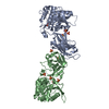
| ||||||||
|---|---|---|---|---|---|---|---|---|---|
| 1 | 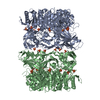
| ||||||||
| 2 | 
| ||||||||
| 3 | 
| ||||||||
| Unit cell |
|
-Components
| #1: Protein | Mass: 43067.750 Da / Num. of mol.: 2 / Fragment: residues 16-395 Source method: isolated from a genetically manipulated source Source: (gene. exp.) Thermotoga maritima (bacteria) / Strain: MSB8 / Gene: TM_0033 / Plasmid: pET-24d / Production host: #2: Chemical | ChemComp-SO4 /   Mass: 96.063 Da / Num. of mol.: 10 / Source method: obtained synthetically / Formula: SO4 Mass: 96.063 Da / Num. of mol.: 10 / Source method: obtained synthetically / Formula: SO4#3: Water | ChemComp-HOH / |  Mass: 18.015 Da / Num. of mol.: 156 / Source method: isolated from a natural source / Formula: H2O Mass: 18.015 Da / Num. of mol.: 156 / Source method: isolated from a natural source / Formula: H2O |
|---|
-Experimental details
-Experiment
| Experiment | Method: X-RAY DIFFRACTION / Number of used crystals: 1 |
|---|
- Sample preparation
| Crystal | Density Matthews: 2.9 Å3/Da / Density % sol: 57.55 % |
|---|---|
| Crystal grow | Temperature: 293 K / Method: vapor diffusion, hanging drop / pH: 7 Details: 20% PEG 8000, 1M lithium sulphate monohydrate, pH 7.0, VAPOR DIFFUSION, HANGING DROP, temperature 293K |
-Data collection
| Diffraction | Mean temperature: 100 K | |||||||||||||||
|---|---|---|---|---|---|---|---|---|---|---|---|---|---|---|---|---|
| Diffraction source | Source: SYNCHROTRON / Site: ESRF  / Beamline: ID29 / Wavelength: 1.0000, 0.9791, 0.9793, 0.9757 / Beamline: ID29 / Wavelength: 1.0000, 0.9791, 0.9793, 0.9757 | |||||||||||||||
| Detector | Type: ADSC QUANTUM 315 / Detector: CCD / Date: Mar 7, 2007 / Details: mirrors | |||||||||||||||
| Radiation | Monochromator: Si(111) monochromator / Protocol: MAD / Monochromatic (M) / Laue (L): M / Scattering type: x-ray | |||||||||||||||
| Radiation wavelength |
| |||||||||||||||
| Reflection | Resolution: 2.6→50 Å / Num. obs: 31079 / % possible obs: 100 % / Observed criterion σ(F): 0 / Observed criterion σ(I): 0 / Redundancy: 5.6 % / Rmerge(I) obs: 0.087 / Net I/σ(I): 14.2 | |||||||||||||||
| Reflection shell | Resolution: 2.6→2.74 Å / Redundancy: 5.7 % / Rmerge(I) obs: 0.43 / Mean I/σ(I) obs: 3.6 / % possible all: 100 |
- Processing
| Software |
| ||||||||||||||||||||||||||||||||||||||||||||||||||||||||||||||||||||||||||||||||||||||||||
|---|---|---|---|---|---|---|---|---|---|---|---|---|---|---|---|---|---|---|---|---|---|---|---|---|---|---|---|---|---|---|---|---|---|---|---|---|---|---|---|---|---|---|---|---|---|---|---|---|---|---|---|---|---|---|---|---|---|---|---|---|---|---|---|---|---|---|---|---|---|---|---|---|---|---|---|---|---|---|---|---|---|---|---|---|---|---|---|---|---|---|---|
| Refinement | Method to determine structure: MAD / Resolution: 2.6→19.96 Å / Cor.coef. Fo:Fc: 0.947 / Cor.coef. Fo:Fc free: 0.898 / SU B: 11.516 / SU ML: 0.245 / Cross valid method: THROUGHOUT / σ(F): 0 / σ(I): 0 / ESU R: 0.693 / ESU R Free: 0.335 / Stereochemistry target values: MAXIMUM LIKELIHOOD
| ||||||||||||||||||||||||||||||||||||||||||||||||||||||||||||||||||||||||||||||||||||||||||
| Solvent computation | Ion probe radii: 0.8 Å / Shrinkage radii: 0.8 Å / VDW probe radii: 1.2 Å / Solvent model: BABINET MODEL WITH MASK | ||||||||||||||||||||||||||||||||||||||||||||||||||||||||||||||||||||||||||||||||||||||||||
| Displacement parameters | Biso mean: 37.122 Å2
| ||||||||||||||||||||||||||||||||||||||||||||||||||||||||||||||||||||||||||||||||||||||||||
| Refinement step | Cycle: LAST / Resolution: 2.6→19.96 Å
| ||||||||||||||||||||||||||||||||||||||||||||||||||||||||||||||||||||||||||||||||||||||||||
| Refine LS restraints |
| ||||||||||||||||||||||||||||||||||||||||||||||||||||||||||||||||||||||||||||||||||||||||||
| LS refinement shell | Resolution: 2.6→2.666 Å / Total num. of bins used: 20
|
