 Movie
Movie Controller

[English] 日本語
 Yorodumi
- PDB-7azl: DNA polymerase sliding clamp from Escherichia coli with peptide 3... -
+ Open data

- Basic information
| Entry | Database: PDB / ID: 7azl | |||||||||
|---|---|---|---|---|---|---|---|---|---|---|
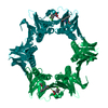
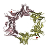
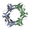
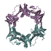
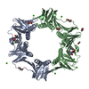
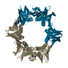
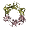
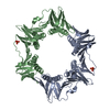
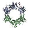
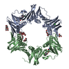
| Title | DNA polymerase sliding clamp from Escherichia coli with peptide 38 bound | |||||||||
 Components Components |
| |||||||||
 Keywords Keywords | DNA BINDING PROTEIN / antibacterial drug | |||||||||
| Function / homology | DNA Polymerase III; Chain A, domain 2 / DNA Polymerase III, subunit A, domain 2 / Roll / Alpha Beta / DI(HYDROXYETHYL)ETHER / :  Function and homology information Function and homology information | |||||||||
| Biological species |  synthetic construct (others) | |||||||||
| Method |  X-RAY DIFFRACTION / SYNCHROTRON / MOLECULAR REPLACEMENT / Resolution: 2.42 Å X-RAY DIFFRACTION / SYNCHROTRON / MOLECULAR REPLACEMENT / Resolution: 2.42 Å | |||||||||
 Authors Authors | Monsarrat, C. / Compain, G. / Andre, C. / Martiel, I. / Engilberge, S. / Olieric, V. / Wolff, P. / Brillet, K. / Landolfo, M. / Silva da Veiga, C. ...Monsarrat, C. / Compain, G. / Andre, C. / Martiel, I. / Engilberge, S. / Olieric, V. / Wolff, P. / Brillet, K. / Landolfo, M. / Silva da Veiga, C. / Wagner, J. / Guichard, G. / Burnouf, D.Y. | |||||||||
 Citation Citation | Journal: J.Med.Chem. / Year: 2021 Title: Iterative Structure-Based Optimization of Short Peptides Targeting the Bacterial Sliding Clamp. Authors: Monsarrat, C. / Compain, G. / Andre, C. / Engilberge, S. / Martiel, I. / Olieric, V. / Wolff, P. / Brillet, K. / Landolfo, M. / Silva da Veiga, C. / Wagner, J. / Guichard, G. / Burnouf, D.Y. | |||||||||
| History |
|
- Structure visualization
| Structure viewer | Molecule: MolmilJmol/JSmol |
|---|
- Downloads & links
-Download
| PDBx/mmCIF format | 7azl.cif.gz | 306.5 KB | Display | PDBx/mmCIF format |
|---|---|---|---|---|
| PDB format | pdb7azl.ent.gz | 243.8 KB | Display | PDB format |
| PDBx/mmJSON format | 7azl.json.gz | Tree view | PDBx/mmJSON format | |
| Others |  Other downloads Other downloads |
-Validation report
| Arichive directory | https://data.pdbj.org/pub/pdb/validation_reports/az/7azlftp://data.pdbj.org/pub/pdb/validation_reports/az/7azl | HTTPS FTP |
|---|
-Related structure data
| Related structure data |  7az5C 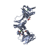 7az6C  7az7C 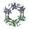 7az8C  7azcC  7azdC 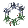 7azeC  7azfC  7azgC  7azkC 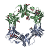 6fvlS S: Starting model for refinement C: citing same article ( |
|---|---|
| Similar structure data |


-Links
 PDBj
PDBj- Assembly
| Deposited unit | 
| ||||||||||||
|---|---|---|---|---|---|---|---|---|---|---|---|---|---|
| 1 | 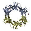
| ||||||||||||
| 2 | 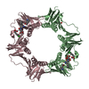
| ||||||||||||
| Unit cell |
|
-Components
| #1: Protein | Mass: 42801.863 Da / Num. of mol.: 4 Source method: isolated from a genetically manipulated source Source: (gene. exp.) Gene: dnaN, AD31_4438 / Production host: #2: Protein/peptide | Mass: 890.849 Da / Num. of mol.: 4 / Source method: obtained synthetically / Source: (synth.) synthetic construct (others) #3: Chemical | ChemComp-GOL / |   Mass: 92.094 Da / Num. of mol.: 1 / Source method: obtained synthetically / Formula: C3H8O3 Mass: 92.094 Da / Num. of mol.: 1 / Source method: obtained synthetically / Formula: C3H8O3#4: Chemical |   Mass: 106.120 Da / Num. of mol.: 2 / Source method: obtained synthetically / Formula: C4H10O3 Mass: 106.120 Da / Num. of mol.: 2 / Source method: obtained synthetically / Formula: C4H10O3#5: Water | ChemComp-HOH / |  Mass: 18.015 Da / Num. of mol.: 417 / Source method: isolated from a natural source / Formula: H2O Mass: 18.015 Da / Num. of mol.: 417 / Source method: isolated from a natural source / Formula: H2OHas ligand of interest | Y | |
|---|
-Experimental details
-Experiment
| Experiment | Method: X-RAY DIFFRACTION / Number of used crystals: 1 |
|---|
- Sample preparation
| Crystal | Density Matthews: 2.72 Å3/Da / Density % sol: 54.71 % |
|---|---|
| Crystal grow | Temperature: 298 K / Method: vapor diffusion / Details: 0.2 M Sodium formate pH 7.2, PEG 3350 20% (w/v) |
-Data collection
| Diffraction | Mean temperature: 100 K / Serial crystal experiment: N |
|---|---|
| Diffraction source | Source: SYNCHROTRON / Site: SLS  / Beamline: X06DA / Wavelength: 1 Å / Beamline: X06DA / Wavelength: 1 Å |
| Detector | Type: DECTRIS PILATUS 2M / Detector: PIXEL / Date: Nov 1, 2018 |
| Radiation | Protocol: SINGLE WAVELENGTH / Monochromatic (M) / Laue (L): M / Scattering type: x-ray |
| Radiation wavelength | Wavelength: 1 Å / Relative weight: 1 |
| Reflection | Resolution: 2.42→70.69 Å / Num. obs: 70023 / % possible obs: 99.9 % / Redundancy: 6.7 % / CC1/2: 0.99 / Rpim(I) all: 0.051 / Net I/σ(I): 11.3 |
| Reflection shell | Resolution: 2.42→2.46 Å / Mean I/σ(I) obs: 2.1 / Num. unique obs: 3408 / CC1/2: 0.72 / Rpim(I) all: 0.39 |
- Processing
| Software |
| ||||||||||||||||||||||||
|---|---|---|---|---|---|---|---|---|---|---|---|---|---|---|---|---|---|---|---|---|---|---|---|---|---|
| Refinement | Method to determine structure: MOLECULAR REPLACEMENT Starting model: 6FVL Resolution: 2.42→70.69 Å / Cross valid method: THROUGHOUT Stereochemistry target values: GeoStd + Monomer Library + CDL v1.2
| ||||||||||||||||||||||||
| Displacement parameters | Biso mean: 49.63 Å2 | ||||||||||||||||||||||||
| Refinement step | Cycle: LAST / Resolution: 2.42→70.69 Å
| ||||||||||||||||||||||||
| Refine LS restraints |
|
