 Movie
Movie Controller

[English] 日本語
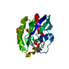
 Yorodumi
- PDB-7aur: Yeast Diphosphoinositol Polyphosphate Phosphohydrolase DDP1 in co... -
+ Open data

- Basic information
| Entry | Database: PDB / ID: 7aur | ||||||
|---|---|---|---|---|---|---|---|
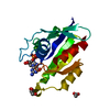
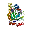
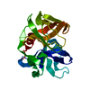
| Title | Yeast Diphosphoinositol Polyphosphate Phosphohydrolase DDP1 in complex with AMP-PNP | ||||||
 Components Components | Diphosphoinositol polyphosphate phosphohydrolase DDP1 | ||||||
 Keywords Keywords | HYDROLASE / Inositol / PP-InsP / Pyrophosphatase / Polyphosphate / Diadenosine polyphosphate / DDP1 / Nudix | ||||||
| Function / homology |  Function and homology information Function and homology informationdiadenosine hexaphosphate hydrolase (AMP-forming) / Synthesis of pyrophosphates in the cytosol / polyphosphate catabolic process / phosphodiesterase decapping endonuclease activity / endopolyphosphatase / diadenosine polyphosphate catabolic process / bis(5'-adenosyl)-hexaphosphatase activity / diadenosine pentaphosphate catabolic process / diadenosine hexaphosphate catabolic process / adenosine 5'-(hexahydrogen pentaphosphate) catabolic process ...diadenosine hexaphosphate hydrolase (AMP-forming) / Synthesis of pyrophosphates in the cytosol / polyphosphate catabolic process / phosphodiesterase decapping endonuclease activity / endopolyphosphatase / diadenosine polyphosphate catabolic process / bis(5'-adenosyl)-hexaphosphatase activity / diadenosine pentaphosphate catabolic process / diadenosine hexaphosphate catabolic process / adenosine 5'-(hexahydrogen pentaphosphate) catabolic process / endopolyphosphatase activity / diphosphoinositol polyphosphate metabolic process / diphosphoinositol-polyphosphate diphosphatase activity / bis(5'-adenosyl)-pentaphosphatase activity / diphosphoinositol-polyphosphate diphosphatase / metal ion binding / nucleus / cytoplasm Similarity search - Function | ||||||
| Biological species |  | ||||||
| Method |  X-RAY DIFFRACTION / SYNCHROTRON / MOLECULAR REPLACEMENT / Resolution: 1.65 Å X-RAY DIFFRACTION / SYNCHROTRON / MOLECULAR REPLACEMENT / Resolution: 1.65 Å | ||||||
 Authors Authors | Marquez-Monino, M.A. / Gonzalez, B. | ||||||
| Funding support |  Spain, 1items Spain, 1items
| ||||||
 Citation Citation | Journal: Sci Adv / Year: 2021 Title: Multiple substrate recognition by yeast diadenosine and diphosphoinositol polyphosphate phosphohydrolase through phosphate clamping. Authors: Marquez-Monino, M.A. / Ortega-Garcia, R. / Shipton, M.L. / Franco-Echevarria, E. / Riley, A.M. / Sanz-Aparicio, J. / Potter, B.V.L. / Gonzalez, B. | ||||||
| History |
|
- Structure visualization
| Structure viewer | Molecule: MolmilJmol/JSmol |
|---|
- Downloads & links
-Download
| PDBx/mmCIF format | 7aur.cif.gz | 51.4 KB | Display | PDBx/mmCIF format |
|---|---|---|---|---|
| PDB format | pdb7aur.ent.gz | 34.7 KB | Display | PDB format |
| PDBx/mmJSON format | 7aur.json.gz | Tree view | PDBx/mmJSON format | |
| Others |  Other downloads Other downloads |
-Validation report
| Arichive directory | https://data.pdbj.org/pub/pdb/validation_reports/au/7aurftp://data.pdbj.org/pub/pdb/validation_reports/au/7aur | HTTPS FTP |
|---|
-Related structure data
| Related structure data | 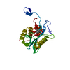 7auiSC 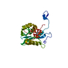 7aujC 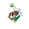 7aukC 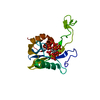 7aulC  7aumC  7aunC  7auoC  7aupC 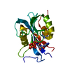 7auqC 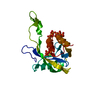 7ausC 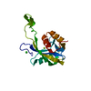 7autC 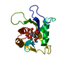 7auuC S: Starting model for refinement C: citing same article ( |
|---|---|
| Similar structure data |


-Links
 PDBj
PDBj


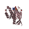
- Assembly
| Deposited unit | 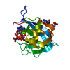
| |||||||||||||||
|---|---|---|---|---|---|---|---|---|---|---|---|---|---|---|---|---|
| 1 |
| |||||||||||||||
| Unit cell |
| |||||||||||||||
| Components on special symmetry positions |
|
-Components
| #1: Protein | Mass: 21812.713 Da / Num. of mol.: 1 Source method: isolated from a genetically manipulated source Details: GGS is a rest of purification linker, protein starts in MGK. Source: (gene. exp.) Strain: ATCC 204508 / S288c / Gene: DDP1, YOR163W, O3575 / Plasmid: pKLSLt / Production host:  References: UniProt: Q99321, diphosphoinositol-polyphosphate diphosphatase, diadenosine hexaphosphate hydrolase (AMP-forming) | ||||||||
|---|---|---|---|---|---|---|---|---|---|
| #2: Chemical |   Mass: 506.196 Da / Num. of mol.: 3 / Source method: obtained synthetically / Formula: C10H17N6O12P3 / Feature type: SUBJECT OF INVESTIGATION / Comment: AMP-PNP, energy-carrying molecule analogue*YM Mass: 506.196 Da / Num. of mol.: 3 / Source method: obtained synthetically / Formula: C10H17N6O12P3 / Feature type: SUBJECT OF INVESTIGATION / Comment: AMP-PNP, energy-carrying molecule analogue*YM#3: Chemical |   Mass: 24.305 Da / Num. of mol.: 2 / Source method: obtained synthetically / Formula: Mg / Feature type: SUBJECT OF INVESTIGATION Mass: 24.305 Da / Num. of mol.: 2 / Source method: obtained synthetically / Formula: Mg / Feature type: SUBJECT OF INVESTIGATION#4: Chemical | ChemComp-CL / |   Mass: 35.453 Da / Num. of mol.: 1 / Source method: obtained synthetically / Formula: Cl Mass: 35.453 Da / Num. of mol.: 1 / Source method: obtained synthetically / Formula: Cl#5: Water | ChemComp-HOH / |  Mass: 18.015 Da / Num. of mol.: 112 / Source method: isolated from a natural source / Formula: H2O Mass: 18.015 Da / Num. of mol.: 112 / Source method: isolated from a natural source / Formula: H2OHas ligand of interest | Y | |
-Experimental details
-Experiment
| Experiment | Method: X-RAY DIFFRACTION / Number of used crystals: 1 |
|---|
- Sample preparation
| Crystal | Density Matthews: 2.44 Å3/Da / Density % sol: 49.64 % / Description: Diamond-like |
|---|---|
| Crystal grow | Temperature: 291 K / Method: vapor diffusion, sitting drop / pH: 5 Details: 25% PEG 6000, 0.1 M Sodium acetate pH 5.0, 0.1 M NaCl. Protein:precipitant ratio 2:1. Protein buffer: 20 mM Tris pH 8.0, 150 mM NaCl, 1 mM DTT, 10 mM AMP-PNP. |
-Data collection
| Diffraction | Mean temperature: 100 K / Serial crystal experiment: N |
|---|---|
| Diffraction source | Source: SYNCHROTRON / Site: ALBA / Beamline: XALOC / Wavelength: 0.9793 Å |
| Detector | Type: DECTRIS PILATUS 6M / Detector: PIXEL / Date: Dec 19, 2018 / Details: KB focusing mirrors |
| Radiation | Monochromator: Si(111) channel-cut, cryocooled / Protocol: SINGLE WAVELENGTH / Monochromatic (M) / Laue (L): M / Scattering type: x-ray |
| Radiation wavelength | Wavelength: 0.9793 Å / Relative weight: 1 |
| Reflection | Resolution: 1.65→47.96 Å / Num. obs: 26240 / % possible obs: 99.8 % / Redundancy: 19 % / Biso Wilson estimate: 27.77 Å2 / CC1/2: 0.999 / Rmerge(I) obs: 0.083 / Rpim(I) all: 0.02 / Net I/σ(I): 18.9 |
| Reflection shell | Resolution: 1.65→1.68 Å / Redundancy: 17.2 % / Rmerge(I) obs: 0.666 / Mean I/σ(I) obs: 4.8 / Num. unique obs: 1299 / CC1/2: 0.937 / Rpim(I) all: 0.164 / % possible all: 99.8 |
- Processing
| Software |
| ||||||||||||||||||||||||||||||||||||||||||||||||||||||||||||
|---|---|---|---|---|---|---|---|---|---|---|---|---|---|---|---|---|---|---|---|---|---|---|---|---|---|---|---|---|---|---|---|---|---|---|---|---|---|---|---|---|---|---|---|---|---|---|---|---|---|---|---|---|---|---|---|---|---|---|---|---|---|
| Refinement | Method to determine structure: MOLECULAR REPLACEMENT Starting model: 7AUI Resolution: 1.65→46.87 Å / Cor.coef. Fo:Fc: 0.963 / Cor.coef. Fo:Fc free: 0.948 / SU B: 2.173 / SU ML: 0.074 / Cross valid method: THROUGHOUT / σ(F): 0 / ESU R: 0.094 / ESU R Free: 0.096 / Stereochemistry target values: MAXIMUM LIKELIHOOD Details: HYDROGENS HAVE BEEN ADDED IN THE RIDING POSITIONS U VALUES : REFINED INDIVIDUALLY
| ||||||||||||||||||||||||||||||||||||||||||||||||||||||||||||
| Solvent computation | Ion probe radii: 0.8 Å / Shrinkage radii: 0.8 Å / VDW probe radii: 1.2 Å / Solvent model: MASK | ||||||||||||||||||||||||||||||||||||||||||||||||||||||||||||
| Displacement parameters | Biso max: 120.48 Å2 / Biso mean: 35.977 Å2 / Biso min: 21.47 Å2
| ||||||||||||||||||||||||||||||||||||||||||||||||||||||||||||
| Refinement step | Cycle: final / Resolution: 1.65→46.87 Å
| ||||||||||||||||||||||||||||||||||||||||||||||||||||||||||||
| Refine LS restraints |
| ||||||||||||||||||||||||||||||||||||||||||||||||||||||||||||
| LS refinement shell | Resolution: 1.65→1.693 Å / Rfactor Rfree error: 0 / Total num. of bins used: 20
|
