 Movie
Movie Controller

[English] 日本語
 Yorodumi
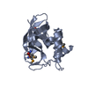
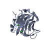
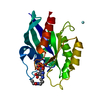
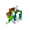
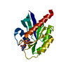
- PDB-2i5t: Crystal Structure of hypothetical protein LOC79017 from Homo sapiens -
+ Open data

- Basic information
| Entry | Database: PDB / ID: 2i5t | ||||||
|---|---|---|---|---|---|---|---|
| Title | Crystal Structure of hypothetical protein LOC79017 from Homo sapiens | ||||||
 Components Components | Protein C7orf24 | ||||||
 Keywords Keywords | STRUCTURAL GENOMICS / UNKNOWN FUNCTION / PSI-2 / Protein Structure Initiative / Center for Eukaryotic Structural Genomics / CESG | ||||||
| Function / homology |  Function and homology information Function and homology informationgamma-glutamylcyclotransferase / gamma-glutamylcyclotransferase activity / Glutathione synthesis and recycling / release of cytochrome c from mitochondria / protein homodimerization activity / extracellular exosome / cytosol Similarity search - Function | ||||||
| Biological species |  Homo sapiens (human) Homo sapiens (human) | ||||||
| Method |  X-RAY DIFFRACTION / SYNCHROTRON / SAD / Resolution: 2.01 Å X-RAY DIFFRACTION / SYNCHROTRON / SAD / Resolution: 2.01 Å | ||||||
 Authors Authors | Bae, E. / Wesenberg, G.E. / Phillips Jr., G.N. / Bitto, E. / Bingman, C.A. / Center for Eukaryotic Structural Genomics (CESG) | ||||||
 Citation Citation | Journal: Proteins / Year: 2008 Title: Crystal structure of Homo sapiens protein LOC79017. Authors: Bae, E. / Bingman, C.A. / Aceti, D.J. / Phillips Jr., G.N. | ||||||
| History |
|
- Structure visualization
| Structure viewer | Molecule: MolmilJmol/JSmol |
|---|
- Downloads & links
-Download
| PDBx/mmCIF format | 2i5t.cif.gz | 85.5 KB | Display | PDBx/mmCIF format |
|---|---|---|---|---|
| PDB format | pdb2i5t.ent.gz | 64.1 KB | Display | PDB format |
| PDBx/mmJSON format | 2i5t.json.gz | Tree view | PDBx/mmJSON format | |
| Others |  Other downloads Other downloads |
-Validation report
| Arichive directory | https://data.pdbj.org/pub/pdb/validation_reports/i5/2i5tftp://data.pdbj.org/pub/pdb/validation_reports/i5/2i5t | HTTPS FTP |
|---|
-Related structure data


-Links
 PDBj
PDBj- Assembly
| Deposited unit | 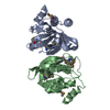
| ||||||||
|---|---|---|---|---|---|---|---|---|---|
| 1 | 
| ||||||||
| 2 | 
| ||||||||
| Unit cell |
| ||||||||
| Details | There are 2 biological units in the asymmetric unit (chains A & B) |
-Components
| #1: Protein | Mass: 21173.152 Da / Num. of mol.: 2 Source method: isolated from a genetically manipulated source Source: (gene. exp.) Homo sapiens (human) / Gene: C7orf24 / Plasmid: PVP 30 / Production host:  #2: Water | ChemComp-HOH / |  Mass: 18.015 Da / Num. of mol.: 141 / Source method: isolated from a natural source / Formula: H2O Mass: 18.015 Da / Num. of mol.: 141 / Source method: isolated from a natural source / Formula: H2OHas protein modification | Y | |
|---|
-Experimental details
-Experiment
| Experiment | Method: X-RAY DIFFRACTION / Number of used crystals: 1 |
|---|
- Sample preparation
| Crystal | Density Matthews: 2.2 Å3/Da / Density % sol: 42.5 % |
|---|---|
| Crystal grow | Temperature: 293 K / Method: vapor diffusion, hanging drop Details: PROTEIN SOLUTION (10 MG/ML PROTEIN, 0.050 M SODIUM CHLORIDE, 0.003 M SODIUM AZIDE, 0.0003 M TCEP, 0.005 M BISTRIS PH 7.0) MIXED IN A 1:1 RATIO WITH THE WELL SOLUTION (28% PEG 2K, 0.20 M ...Details: PROTEIN SOLUTION (10 MG/ML PROTEIN, 0.050 M SODIUM CHLORIDE, 0.003 M SODIUM AZIDE, 0.0003 M TCEP, 0.005 M BISTRIS PH 7.0) MIXED IN A 1:1 RATIO WITH THE WELL SOLUTION (28% PEG 2K, 0.20 M POTASSIUM GLUTAMATE, 0.1 M MES/ACETATE PH 5.5) CRYOPROTECTED WITH 10% ETHYLENE GLYCOL IN WELL SOLUTION, VAPOR DIFFUSION, HANGING DROP, temperature 293K |
-Data collection
| Diffraction | Mean temperature: 100 K |
|---|---|
| Diffraction source | Source: SYNCHROTRON / Site: APS  / Beamline: 23-ID-D / Wavelength: 0.9793 / Beamline: 23-ID-D / Wavelength: 0.9793 |
| Detector | Type: MARMOSAIC 300 mm CCD / Detector: CCD / Date: Jun 18, 2006 / Details: ADJUSTABLE FOCUSING MIRRORS IN K-B GEOMETRY |
| Radiation | Monochromator: SI(111) DOUBLE CRYSTAL MONOCHROMATER / Protocol: SAD / Monochromatic (M) / Laue (L): M / Scattering type: x-ray |
| Radiation wavelength | Wavelength: 0.9793 Å / Relative weight: 1 |
| Reflection | Resolution: 2.01→45.621 Å / Num. obs: 24297 / % possible obs: 97.5 % / Redundancy: 10.8 % / Rmerge(I) obs: 0.123 / Net I/σ(I): 12.185 |
| Reflection shell | Resolution: 2.01→2.08 Å / Redundancy: 8.1 % / Rmerge(I) obs: 0.357 / Mean I/σ(I) obs: 4.096 / % possible all: 83.2 |
-Phasing
| Phasing | Method: SAD | ||||||||||||||||||||||||||||||||||||||||||||||||||||||||||||||||||||||||||||||||||||||||||||||||||||||||||||||||||||||||||||||||||||||||||||||||||||||||||||||||||||||||||||||||||||||||||||||||||||||||||||||||||||||||||||||||||||||||||||||||||||||
|---|---|---|---|---|---|---|---|---|---|---|---|---|---|---|---|---|---|---|---|---|---|---|---|---|---|---|---|---|---|---|---|---|---|---|---|---|---|---|---|---|---|---|---|---|---|---|---|---|---|---|---|---|---|---|---|---|---|---|---|---|---|---|---|---|---|---|---|---|---|---|---|---|---|---|---|---|---|---|---|---|---|---|---|---|---|---|---|---|---|---|---|---|---|---|---|---|---|---|---|---|---|---|---|---|---|---|---|---|---|---|---|---|---|---|---|---|---|---|---|---|---|---|---|---|---|---|---|---|---|---|---|---|---|---|---|---|---|---|---|---|---|---|---|---|---|---|---|---|---|---|---|---|---|---|---|---|---|---|---|---|---|---|---|---|---|---|---|---|---|---|---|---|---|---|---|---|---|---|---|---|---|---|---|---|---|---|---|---|---|---|---|---|---|---|---|---|---|---|---|---|---|---|---|---|---|---|---|---|---|---|---|---|---|---|---|---|---|---|---|---|---|---|---|---|---|---|---|---|---|---|---|---|---|---|---|---|---|---|---|---|---|---|---|---|---|---|---|
| Phasing MAD set | R cullis centric: 0 / Highest resolution: 2.01 Å / Lowest resolution: 44.99 Å / Power centric: 0
| ||||||||||||||||||||||||||||||||||||||||||||||||||||||||||||||||||||||||||||||||||||||||||||||||||||||||||||||||||||||||||||||||||||||||||||||||||||||||||||||||||||||||||||||||||||||||||||||||||||||||||||||||||||||||||||||||||||||||||||||||||||||
| Phasing MAD set shell | R cullis centric: 0 / Power centric: 0
| ||||||||||||||||||||||||||||||||||||||||||||||||||||||||||||||||||||||||||||||||||||||||||||||||||||||||||||||||||||||||||||||||||||||||||||||||||||||||||||||||||||||||||||||||||||||||||||||||||||||||||||||||||||||||||||||||||||||||||||||||||||||
| Phasing MAD set site |
| ||||||||||||||||||||||||||||||||||||||||||||||||||||||||||||||||||||||||||||||||||||||||||||||||||||||||||||||||||||||||||||||||||||||||||||||||||||||||||||||||||||||||||||||||||||||||||||||||||||||||||||||||||||||||||||||||||||||||||||||||||||||
| Phasing dm | Method: Solvent flattening and Histogram matching / Reflection: 24211 | ||||||||||||||||||||||||||||||||||||||||||||||||||||||||||||||||||||||||||||||||||||||||||||||||||||||||||||||||||||||||||||||||||||||||||||||||||||||||||||||||||||||||||||||||||||||||||||||||||||||||||||||||||||||||||||||||||||||||||||||||||||||
| Phasing dm shell |
|
- Processing
| Software |
| ||||||||||||||||||||||||||||||||||||||||||||||||||||||||||||||||||||||||||||||||||||||||||||||||||||||||||||||||||||||||||||||||||||||||||||||||||||||||||||||||||||||||||
|---|---|---|---|---|---|---|---|---|---|---|---|---|---|---|---|---|---|---|---|---|---|---|---|---|---|---|---|---|---|---|---|---|---|---|---|---|---|---|---|---|---|---|---|---|---|---|---|---|---|---|---|---|---|---|---|---|---|---|---|---|---|---|---|---|---|---|---|---|---|---|---|---|---|---|---|---|---|---|---|---|---|---|---|---|---|---|---|---|---|---|---|---|---|---|---|---|---|---|---|---|---|---|---|---|---|---|---|---|---|---|---|---|---|---|---|---|---|---|---|---|---|---|---|---|---|---|---|---|---|---|---|---|---|---|---|---|---|---|---|---|---|---|---|---|---|---|---|---|---|---|---|---|---|---|---|---|---|---|---|---|---|---|---|---|---|---|---|---|---|---|---|
| Refinement | Method to determine structure: SAD / Resolution: 2.01→45.62 Å / Cor.coef. Fo:Fc: 0.946 / Cor.coef. Fo:Fc free: 0.918 / SU B: 4.02 / SU ML: 0.114 / Cross valid method: THROUGHOUT / σ(F): 0 / ESU R: 0.189 / ESU R Free: 0.171 / Stereochemistry target values: MAXIMUM LIKELIHOOD Details: HYDROGENS HAVE BEEN ADDED IN THE RIDING POSITIONS. THE FINAL DIFFERENCE ELECTRON DENSITY MAP SHOWS EXTENSIVE POSITIVE ELECTRON DENSITY WITHIN THE REGION SURROUNDED BY RESIDUES TYR 19, TYR ...Details: HYDROGENS HAVE BEEN ADDED IN THE RIDING POSITIONS. THE FINAL DIFFERENCE ELECTRON DENSITY MAP SHOWS EXTENSIVE POSITIVE ELECTRON DENSITY WITHIN THE REGION SURROUNDED BY RESIDUES TYR 19, TYR 22, GLY 23, SER 24, ASN 25, GLU 98, TYR 105 AND TYR 139. ATTEMPTS TO MODEL THIS DENSITY WITH CRYSTALLIZATION SOLUTION COMPONENTS WERE UNSUCCESSFUL. THE DEPOSITED STRUCTURE FACTOR FILE MAY BE USED TO EXAMINE THIS DENSITY.
| ||||||||||||||||||||||||||||||||||||||||||||||||||||||||||||||||||||||||||||||||||||||||||||||||||||||||||||||||||||||||||||||||||||||||||||||||||||||||||||||||||||||||||
| Solvent computation | Ion probe radii: 0.8 Å / Shrinkage radii: 0.8 Å / VDW probe radii: 1.2 Å / Solvent model: BABINET MODEL PLUS MASK | ||||||||||||||||||||||||||||||||||||||||||||||||||||||||||||||||||||||||||||||||||||||||||||||||||||||||||||||||||||||||||||||||||||||||||||||||||||||||||||||||||||||||||
| Displacement parameters | Biso mean: 23.54 Å2
| ||||||||||||||||||||||||||||||||||||||||||||||||||||||||||||||||||||||||||||||||||||||||||||||||||||||||||||||||||||||||||||||||||||||||||||||||||||||||||||||||||||||||||
| Refinement step | Cycle: LAST / Resolution: 2.01→45.62 Å
| ||||||||||||||||||||||||||||||||||||||||||||||||||||||||||||||||||||||||||||||||||||||||||||||||||||||||||||||||||||||||||||||||||||||||||||||||||||||||||||||||||||||||||
| Refine LS restraints |
| ||||||||||||||||||||||||||||||||||||||||||||||||||||||||||||||||||||||||||||||||||||||||||||||||||||||||||||||||||||||||||||||||||||||||||||||||||||||||||||||||||||||||||
| LS refinement shell | Resolution: 2.01→2.06 Å / Total num. of bins used: 20
|
