 Movie
Movie Controller

[English] 日本語
 Yorodumi
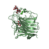
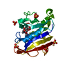
- PDB-3lbf: Crystal structure of Protein L-isoaspartyl methyltransferase from... -
+ Open data

- Basic information
| Entry | Database: PDB / ID: 3lbf | ||||||
|---|---|---|---|---|---|---|---|
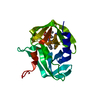
| Title | Crystal structure of Protein L-isoaspartyl methyltransferase from Escherichia coli | ||||||
 Components Components | Protein-L-isoaspartate O-methyltransferase | ||||||
 Keywords Keywords | TRANSFERASE / modified Rossman-type fold / Methyltransferase / S-adenosyl-L-methionine | ||||||
| Function / homology |  Function and homology information Function and homology informationprotein-L-isoaspartate(D-aspartate) O-methyltransferase / protein-L-isoaspartate (D-aspartate) O-methyltransferase activity / protein repair / methylation / cytoplasm Similarity search - Function | ||||||
| Biological species |  | ||||||
| Method |  X-RAY DIFFRACTION / MOLECULAR REPLACEMENT / molecular replacement / Resolution: 1.8 Å X-RAY DIFFRACTION / MOLECULAR REPLACEMENT / molecular replacement / Resolution: 1.8 Å | ||||||
 Authors Authors | Fang, P. / Li, X. / Wang, J. / Niu, L. / Teng, M. | ||||||
 Citation Citation | Journal: Cell Biochem.Biophys. / Year: 2010 Title: Crystal structure of the protein L-isoaspartyl methyltransferase from Escherichia coli Authors: Fang, P. / Li, X. / Wang, J. / Xing, L. / Gao, Y. / Niu, L. / Teng, M. | ||||||
| History |
|
- Structure visualization
| Structure viewer | Molecule: MolmilJmol/JSmol |
|---|
- Downloads & links
-Download
| PDBx/mmCIF format | 3lbf.cif.gz | 335.2 KB | Display | PDBx/mmCIF format |
|---|---|---|---|---|
| PDB format | pdb3lbf.ent.gz | 271.6 KB | Display | PDB format |
| PDBx/mmJSON format | 3lbf.json.gz | Tree view | PDBx/mmJSON format | |
| Others |  Other downloads Other downloads |
-Validation report
| Arichive directory | https://data.pdbj.org/pub/pdb/validation_reports/lb/3lbfftp://data.pdbj.org/pub/pdb/validation_reports/lb/3lbf | HTTPS FTP |
|---|
-Related structure data
| Related structure data | 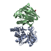 2yxeS S: Starting model for refinement |
|---|---|
| Similar structure data |


-Links
 PDBj
PDBj- Assembly
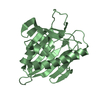
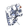
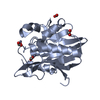
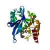
| Deposited unit | 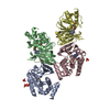
| ||||||||
|---|---|---|---|---|---|---|---|---|---|
| 1 | 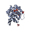
| ||||||||
| 2 | 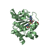
| ||||||||
| 3 | 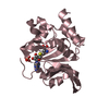
| ||||||||
| 4 | 
| ||||||||
| Unit cell |
|
-Components
| #1: Protein | Mass: 23485.971 Da / Num. of mol.: 4 Source method: isolated from a genetically manipulated source Source: (gene. exp.) References: UniProt: P0A7A5, protein-L-isoaspartate(D-aspartate) O-methyltransferase #2: Chemical | ChemComp-SAH /   Type: L-peptide linking / Mass: 384.411 Da / Num. of mol.: 4 / Source method: obtained synthetically / Formula: C14H20N6O5S Type: L-peptide linking / Mass: 384.411 Da / Num. of mol.: 4 / Source method: obtained synthetically / Formula: C14H20N6O5S#3: Chemical | ChemComp-PO4 /   Mass: 94.971 Da / Num. of mol.: 6 / Source method: obtained synthetically / Formula: PO4 Mass: 94.971 Da / Num. of mol.: 6 / Source method: obtained synthetically / Formula: PO4#4: Chemical | ChemComp-GOL / |   Mass: 92.094 Da / Num. of mol.: 1 / Source method: obtained synthetically / Formula: C3H8O3 Mass: 92.094 Da / Num. of mol.: 1 / Source method: obtained synthetically / Formula: C3H8O3#5: Water | ChemComp-HOH / |  Mass: 18.015 Da / Num. of mol.: 360 / Source method: isolated from a natural source / Formula: H2O Mass: 18.015 Da / Num. of mol.: 360 / Source method: isolated from a natural source / Formula: H2O |
|---|
-Experimental details
-Experiment
| Experiment | Method: X-RAY DIFFRACTION / Number of used crystals: 1 |
|---|
- Sample preparation
| Crystal | Density Matthews: 1.92 Å3/Da / Density % sol: 35.87 % |
|---|---|
| Crystal grow | Temperature: 288 K / Method: vapor diffusion, hanging drop / pH: 7.5 Details: Hepes-Na, monosodium dihydrogen phosphate, monopotassium dihydrogen phosphate, pH 7.5, VAPOR DIFFUSION, HANGING DROP, temperature 288K |
-Data collection
| Diffraction | Mean temperature: 100 K |
|---|---|
| Diffraction source | Source: ROTATING ANODE / Type: RIGAKU MICROMAX-007 / Wavelength: 1 Å |
| Detector | Date: Oct 28, 2009 |
| Radiation | Protocol: SINGLE WAVELENGTH / Monochromatic (M) / Laue (L): M / Scattering type: x-ray |
| Radiation wavelength | Wavelength: 1 Å / Relative weight: 1 |
| Reflection | Resolution: 1.8→25 Å / Num. obs: 62259 / % possible obs: 96.2 % / Redundancy: 2.8 % / Rmerge(I) obs: 0.056 / Net I/σ(I): 10.7 |
| Reflection shell | Resolution: 1.8→1.9 Å / % possible obs: 95 % / Redundancy: 2.8 % / Rmerge(I) obs: 0.207 / Mean I/σ(I) obs: 3.9 / Num. unique all: 8977 |
-Phasing
| Phasing | Method: molecular replacement |
|---|
- Processing
| Software |
| ||||||||||||||||||||||||||||||||||||||||||||||||||||||||||||||||||||||||||||||||||||||||||||||||||||||||||||||||||||||||||||||||||||||||||||||||||||||||||||||||||||||||||||||||||||||||||||||||||||||||
|---|---|---|---|---|---|---|---|---|---|---|---|---|---|---|---|---|---|---|---|---|---|---|---|---|---|---|---|---|---|---|---|---|---|---|---|---|---|---|---|---|---|---|---|---|---|---|---|---|---|---|---|---|---|---|---|---|---|---|---|---|---|---|---|---|---|---|---|---|---|---|---|---|---|---|---|---|---|---|---|---|---|---|---|---|---|---|---|---|---|---|---|---|---|---|---|---|---|---|---|---|---|---|---|---|---|---|---|---|---|---|---|---|---|---|---|---|---|---|---|---|---|---|---|---|---|---|---|---|---|---|---|---|---|---|---|---|---|---|---|---|---|---|---|---|---|---|---|---|---|---|---|---|---|---|---|---|---|---|---|---|---|---|---|---|---|---|---|---|---|---|---|---|---|---|---|---|---|---|---|---|---|---|---|---|---|---|---|---|---|---|---|---|---|---|---|---|---|---|---|---|---|
| Refinement | Method to determine structure: MOLECULAR REPLACEMENT Starting model: 2YXE Resolution: 1.8→25 Å / Cor.coef. Fo:Fc: 0.952 / Cor.coef. Fo:Fc free: 0.935 / Occupancy max: 1 / Occupancy min: 0.15 / Cross valid method: THROUGHOUT / σ(F): 0 / ESU R: 0.179 / ESU R Free: 0.154 / Stereochemistry target values: MAXIMUM LIKELIHOOD Details: HYDROGENS HAVE BEEN ADDED IN THE RIDING POSITIONS U VALUES: WITH TLS ADDED
| ||||||||||||||||||||||||||||||||||||||||||||||||||||||||||||||||||||||||||||||||||||||||||||||||||||||||||||||||||||||||||||||||||||||||||||||||||||||||||||||||||||||||||||||||||||||||||||||||||||||||
| Solvent computation | Ion probe radii: 0.8 Å / Shrinkage radii: 0.8 Å / VDW probe radii: 1.4 Å / Solvent model: MASK | ||||||||||||||||||||||||||||||||||||||||||||||||||||||||||||||||||||||||||||||||||||||||||||||||||||||||||||||||||||||||||||||||||||||||||||||||||||||||||||||||||||||||||||||||||||||||||||||||||||||||
| Displacement parameters | Biso max: 222.56 Å2 / Biso mean: 42.2995 Å2 / Biso min: 16.42 Å2
| ||||||||||||||||||||||||||||||||||||||||||||||||||||||||||||||||||||||||||||||||||||||||||||||||||||||||||||||||||||||||||||||||||||||||||||||||||||||||||||||||||||||||||||||||||||||||||||||||||||||||
| Refinement step | Cycle: LAST / Resolution: 1.8→25 Å
| ||||||||||||||||||||||||||||||||||||||||||||||||||||||||||||||||||||||||||||||||||||||||||||||||||||||||||||||||||||||||||||||||||||||||||||||||||||||||||||||||||||||||||||||||||||||||||||||||||||||||
| Refine LS restraints |
| ||||||||||||||||||||||||||||||||||||||||||||||||||||||||||||||||||||||||||||||||||||||||||||||||||||||||||||||||||||||||||||||||||||||||||||||||||||||||||||||||||||||||||||||||||||||||||||||||||||||||
| LS refinement shell | Resolution: 1.8→1.847 Å / Total num. of bins used: 20
| ||||||||||||||||||||||||||||||||||||||||||||||||||||||||||||||||||||||||||||||||||||||||||||||||||||||||||||||||||||||||||||||||||||||||||||||||||||||||||||||||||||||||||||||||||||||||||||||||||||||||
| Refinement TLS params. | Method: refined / Refine-ID: X-RAY DIFFRACTION
| ||||||||||||||||||||||||||||||||||||||||||||||||||||||||||||||||||||||||||||||||||||||||||||||||||||||||||||||||||||||||||||||||||||||||||||||||||||||||||||||||||||||||||||||||||||||||||||||||||||||||
| Refinement TLS group |
|
