 Movie
Movie Controller

[English] 日本語
 Yorodumi
- PDB-3ofk: Crystal structure of N-methyltransferase NodS from Bradyrhizobium... -
+ Open data

- Basic information
| Entry | Database: PDB / ID: 3ofk | ||||||
|---|---|---|---|---|---|---|---|
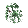
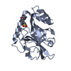
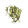
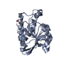
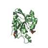
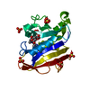
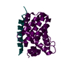
| Title | Crystal structure of N-methyltransferase NodS from Bradyrhizobium japonicum WM9 in complex with S-adenosyl-l-homocysteine (SAH) | ||||||
 Components Components | Nodulation protein S | ||||||
 Keywords Keywords | TRANSFERASE / NODS / N-METHYLTRANSFERASE / SAH / SAM / NOD FACTOR / NODULATION / NITROGEN FIXATION / SYMBIOSIS / alpha/beta structure / variant of Rossmann fold / SAM-dependent N-methyltransferase / S-adenosyl-L-methionine (SAM) / lipochitooligosaccharide / Methylation | ||||||
| Function / homology |  Function and homology information Function and homology informationoligosaccharide biosynthetic process / S-adenosylmethionine-dependent methyltransferase activity / methylation Similarity search - Function | ||||||
| Biological species |  Bradyrhizobium sp. (bacteria) Bradyrhizobium sp. (bacteria) | ||||||
| Method |  X-RAY DIFFRACTION / SYNCHROTRON / SAD / Resolution: 1.85 Å X-RAY DIFFRACTION / SYNCHROTRON / SAD / Resolution: 1.85 Å | ||||||
 Authors Authors | Cakici, O. / Sikorski, M. / Stepkowski, T. / Bujacz, G. / Jaskolski, M. | ||||||
 Citation Citation | Journal: J.Mol.Biol. / Year: 2010 Title: Crystal Structures of NodS N-Methyltransferase from Bradyrhizobium japonicum in Ligand-Free Form and as SAH Complex. Authors: Cakici, O. / Sikorski, M. / Stepkowski, T. / Bujacz, G. / Jaskolski, M. #1: Journal: Acta Crystallogr.,Sect.F / Year: 2008 Title: Cloning, expression, purification, crystallization and preliminary X-ray analysis of NodS N-methyltransferase from Bradyrhizobium japonicum WM9 Authors: Cakici, O. / Sikorski, M. / Stepkowski, T. / Bujacz, G. / Jaskolski, M. | ||||||
| History |
|
- Structure visualization
| Structure viewer | Molecule: MolmilJmol/JSmol |
|---|
- Downloads & links
-Download
| PDBx/mmCIF format | 3ofk.cif.gz | 187.5 KB | Display | PDBx/mmCIF format |
|---|---|---|---|---|
| PDB format | pdb3ofk.ent.gz | 148.8 KB | Display | PDB format |
| PDBx/mmJSON format | 3ofk.json.gz | Tree view | PDBx/mmJSON format | |
| Others |  Other downloads Other downloads |
-Validation report
| Arichive directory | https://data.pdbj.org/pub/pdb/validation_reports/of/3ofkftp://data.pdbj.org/pub/pdb/validation_reports/of/3ofk | HTTPS FTP |
|---|
-Related structure data


-Links
 PDBj
PDBj- Assembly
| Deposited unit | 
| ||||||||
|---|---|---|---|---|---|---|---|---|---|
| 1 | 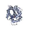
| ||||||||
| 2 | 
| ||||||||
| 3 | 
| ||||||||
| 4 | 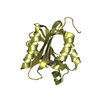
| ||||||||
| Unit cell |
|
-Components
| #1: Protein | Mass: 24152.400 Da / Num. of mol.: 4 Source method: isolated from a genetically manipulated source Details: The crystallized protein has a heptapeptide tag (GIDPFTM-) attached at the N-terminus (residues -6 through 0). The first codon of the nodS sequence (gtg) has been translated in this NodS ...Details: The crystallized protein has a heptapeptide tag (GIDPFTM-) attached at the N-terminus (residues -6 through 0). The first codon of the nodS sequence (gtg) has been translated in this NodS protein as Val1, which is different from the Met1 residue occurring in NodS isolated from Bradyrhizobium japonicum WM9. The present protein is therefore a single-site variant of the natural polypeptide. Source: (gene. exp.) Bradyrhizobium sp. (bacteria) / Strain: WM9 / Gene: nodS / Plasmid: pET151/D-TOPO / Production host: References: UniProt: Q9AQ22, Transferases; Transferring one-carbon groups; Methyltransferases #2: Chemical | ChemComp-SAH /   Type: L-peptide linking / Mass: 384.411 Da / Num. of mol.: 4 / Source method: obtained synthetically / Formula: C14H20N6O5S Type: L-peptide linking / Mass: 384.411 Da / Num. of mol.: 4 / Source method: obtained synthetically / Formula: C14H20N6O5S#3: Water | ChemComp-HOH / |  Mass: 18.015 Da / Num. of mol.: 846 / Source method: isolated from a natural source / Formula: H2O Mass: 18.015 Da / Num. of mol.: 846 / Source method: isolated from a natural source / Formula: H2OSequence details | THE AUTHORS STATE THAT VAL1 IS THE FIRST RESIDUE OF THE PROTEIN SEQUENCE AND CORRESPONDS TO ...THE AUTHORS STATE THAT VAL1 IS THE FIRST RESIDUE OF THE PROTEIN SEQUENCE AND CORRESPOND | |
|---|
-Experimental details
-Experiment
| Experiment | Method: X-RAY DIFFRACTION / Number of used crystals: 1 |
|---|
- Sample preparation
| Crystal | Density Matthews: 2.28 Å3/Da / Density % sol: 46.02 % |
|---|---|
| Crystal grow | Temperature: 292 K / Method: vapor diffusion, hanging drop / pH: 8.5 Details: 16% PEG 8000, 5 mM magnesium chloride, pH 8.5, VAPOR DIFFUSION, HANGING DROP, temperature 292K |
-Data collection
| Diffraction | Mean temperature: 100 K |
|---|---|
| Diffraction source | Source: SYNCHROTRON / Site: BESSY  / Beamline: 14.2 / Wavelength: 0.9184 Å / Beamline: 14.2 / Wavelength: 0.9184 Å |
| Detector | Type: MAR CCD 165 mm / Detector: CCD / Date: Mar 19, 2008 / Details: mirrors |
| Radiation | Monochromator: Si(111) / Protocol: SINGLE WAVELENGTH / Monochromatic (M) / Laue (L): M / Scattering type: x-ray |
| Radiation wavelength | Wavelength: 0.9184 Å / Relative weight: 1 |
| Reflection | Resolution: 1.85→50 Å / Num. obs: 72827 / % possible obs: 96 % / Observed criterion σ(I): -3 / Redundancy: 5.04 % / Biso Wilson estimate: 39 Å2 / Rmerge(I) obs: 0.052 / Net I/σ(I): 24.6 |
| Reflection shell | Resolution: 1.85→1.92 Å / Rmerge(I) obs: 0.347 / Mean I/σ(I) obs: 2.4 / % possible all: 97.8 |
- Processing
| Software |
| |||||||||||||||||||||||||||||||||||||||||||||||||||||||||||||||||||||||||||||||||||||||||||||||||||||||||||||||||||||||||||||||||||||||||||||||||||||||||||||||||||||||||||||||||||||||||||||||||||||||||||||||||||||||||||||||||
|---|---|---|---|---|---|---|---|---|---|---|---|---|---|---|---|---|---|---|---|---|---|---|---|---|---|---|---|---|---|---|---|---|---|---|---|---|---|---|---|---|---|---|---|---|---|---|---|---|---|---|---|---|---|---|---|---|---|---|---|---|---|---|---|---|---|---|---|---|---|---|---|---|---|---|---|---|---|---|---|---|---|---|---|---|---|---|---|---|---|---|---|---|---|---|---|---|---|---|---|---|---|---|---|---|---|---|---|---|---|---|---|---|---|---|---|---|---|---|---|---|---|---|---|---|---|---|---|---|---|---|---|---|---|---|---|---|---|---|---|---|---|---|---|---|---|---|---|---|---|---|---|---|---|---|---|---|---|---|---|---|---|---|---|---|---|---|---|---|---|---|---|---|---|---|---|---|---|---|---|---|---|---|---|---|---|---|---|---|---|---|---|---|---|---|---|---|---|---|---|---|---|---|---|---|---|---|---|---|---|---|---|---|---|---|---|---|---|---|---|---|---|---|---|---|---|---|
| Refinement | Method to determine structure: SAD / Resolution: 1.85→29.7 Å / Cor.coef. Fo:Fc: 0.968 / Cor.coef. Fo:Fc free: 0.95 / SU B: 9.331 / SU ML: 0.127 / Cross valid method: R-FREE / ESU R: 0.145 / ESU R Free: 0.14 / Stereochemistry target values: MAXIMUM LIKELIHOOD / Details: HYDROGEN ATOMS WERE ADDED AT RIDING POSITIONS
| |||||||||||||||||||||||||||||||||||||||||||||||||||||||||||||||||||||||||||||||||||||||||||||||||||||||||||||||||||||||||||||||||||||||||||||||||||||||||||||||||||||||||||||||||||||||||||||||||||||||||||||||||||||||||||||||||
| Solvent computation | Ion probe radii: 0.8 Å / Shrinkage radii: 0.8 Å / VDW probe radii: 1.4 Å / Solvent model: BABINET MODEL WITH MASK | |||||||||||||||||||||||||||||||||||||||||||||||||||||||||||||||||||||||||||||||||||||||||||||||||||||||||||||||||||||||||||||||||||||||||||||||||||||||||||||||||||||||||||||||||||||||||||||||||||||||||||||||||||||||||||||||||
| Displacement parameters | Biso mean: 39.766 Å2
| |||||||||||||||||||||||||||||||||||||||||||||||||||||||||||||||||||||||||||||||||||||||||||||||||||||||||||||||||||||||||||||||||||||||||||||||||||||||||||||||||||||||||||||||||||||||||||||||||||||||||||||||||||||||||||||||||
| Refinement step | Cycle: LAST / Resolution: 1.85→29.7 Å
| |||||||||||||||||||||||||||||||||||||||||||||||||||||||||||||||||||||||||||||||||||||||||||||||||||||||||||||||||||||||||||||||||||||||||||||||||||||||||||||||||||||||||||||||||||||||||||||||||||||||||||||||||||||||||||||||||
| Refine LS restraints |
| |||||||||||||||||||||||||||||||||||||||||||||||||||||||||||||||||||||||||||||||||||||||||||||||||||||||||||||||||||||||||||||||||||||||||||||||||||||||||||||||||||||||||||||||||||||||||||||||||||||||||||||||||||||||||||||||||
| LS refinement shell | Resolution: 1.851→1.899 Å / Total num. of bins used: 20
| |||||||||||||||||||||||||||||||||||||||||||||||||||||||||||||||||||||||||||||||||||||||||||||||||||||||||||||||||||||||||||||||||||||||||||||||||||||||||||||||||||||||||||||||||||||||||||||||||||||||||||||||||||||||||||||||||
| Refinement TLS params. | Method: refined / Refine-ID: X-RAY DIFFRACTION
| |||||||||||||||||||||||||||||||||||||||||||||||||||||||||||||||||||||||||||||||||||||||||||||||||||||||||||||||||||||||||||||||||||||||||||||||||||||||||||||||||||||||||||||||||||||||||||||||||||||||||||||||||||||||||||||||||
| Refinement TLS group |
|
