 Movie
Movie Controller

[English] 日本語
 Yorodumi
- PDB-1jss: Crystal structure of the Mus musculus cholesterol-regulated START... -
+ Open data

- Basic information
| Entry | Database: PDB / ID: 1jss | ||||||
|---|---|---|---|---|---|---|---|
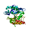
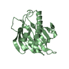
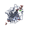
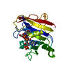
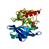
| Title | Crystal structure of the Mus musculus cholesterol-regulated START protein 4 (StarD4). | ||||||
 Components Components | cholesterol-regulated START protein 4 | ||||||
 Keywords Keywords | LIPID BINDING PROTEIN / START domain / Structural Genomics / PSI / Protein Structure Initiative / New York SGX Research Center for Structural Genomics / NYSGXRC | ||||||
| Function / homology |  Function and homology information Function and homology informationcholesterol transport involved in cholesterol storage / Pregnenolone biosynthesis / positive regulation of bile acid biosynthetic process / intracellular cholesterol transport / cholesterol import / positive regulation of cholesterol metabolic process / cholesterol transfer activity / cholesterol binding / cytoplasmic vesicle / endoplasmic reticulum ...cholesterol transport involved in cholesterol storage / Pregnenolone biosynthesis / positive regulation of bile acid biosynthetic process / intracellular cholesterol transport / cholesterol import / positive regulation of cholesterol metabolic process / cholesterol transfer activity / cholesterol binding / cytoplasmic vesicle / endoplasmic reticulum / mitochondrion / cytosol / cytoplasm Similarity search - Function | ||||||
| Biological species |  | ||||||
| Method |  X-RAY DIFFRACTION / SYNCHROTRON / MAD / Resolution: 2.2 Å X-RAY DIFFRACTION / SYNCHROTRON / MAD / Resolution: 2.2 Å | ||||||
 Authors Authors | Romanowski, M.J. / Soccio, R.E. / Breslow, J.L. / Burley, S.K. / New York SGX Research Center for Structural Genomics (NYSGXRC) | ||||||
 Citation Citation | Journal: Proc.Natl.Acad.Sci.USA / Year: 2002 Title: Crystal structure of the Mus musculus cholesterol-regulated START protein 4 (StarD4) containing a StAR-related lipid transfer domain. Authors: Romanowski, M.J. / Soccio, R.E. / Breslow, J.L. / Burley, S.K. | ||||||
| History |
|
- Structure visualization
| Structure viewer | Molecule: MolmilJmol/JSmol |
|---|
- Downloads & links
-Download
| PDBx/mmCIF format | 1jss.cif.gz | 96.9 KB | Display | PDBx/mmCIF format |
|---|---|---|---|---|
| PDB format | pdb1jss.ent.gz | 74.7 KB | Display | PDB format |
| PDBx/mmJSON format | 1jss.json.gz | Tree view | PDBx/mmJSON format | |
| Others |  Other downloads Other downloads |
-Validation report
| Arichive directory | https://data.pdbj.org/pub/pdb/validation_reports/js/1jssftp://data.pdbj.org/pub/pdb/validation_reports/js/1jss | HTTPS FTP |
|---|
-Related structure data
| Similar structure data | |
|---|---|
| Other databases |


-Links
 PDBj
PDBj- Assembly
| Deposited unit | 
| ||||||||
|---|---|---|---|---|---|---|---|---|---|
| 1 | 
| ||||||||
| 2 | 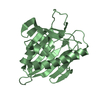
| ||||||||
| Unit cell |
|
-Components
| #1: Protein | Mass: 25579.947 Da / Num. of mol.: 2 / Mutation: K41E, V51A Source method: isolated from a genetically manipulated source Details: similar to RIKEN cDNA 2310058G22 gene (Mus musculus) Source: (gene. exp.)  #2: Water | ChemComp-HOH / |  Mass: 18.015 Da / Num. of mol.: 290 / Source method: isolated from a natural source / Formula: H2O Mass: 18.015 Da / Num. of mol.: 290 / Source method: isolated from a natural source / Formula: H2O |
|---|
-Experimental details
-Experiment
| Experiment | Method: X-RAY DIFFRACTION / Number of used crystals: 1 |
|---|
- Sample preparation
| Crystal | Density Matthews: 2.35 Å3/Da / Density % sol: 47.71 % | |||||||||||||||||||||||||||||||||||||||||||||||||||||||||||||||
|---|---|---|---|---|---|---|---|---|---|---|---|---|---|---|---|---|---|---|---|---|---|---|---|---|---|---|---|---|---|---|---|---|---|---|---|---|---|---|---|---|---|---|---|---|---|---|---|---|---|---|---|---|---|---|---|---|---|---|---|---|---|---|---|---|
| Crystal grow | Temperature: 277 K / Method: vapor diffusion, sitting drop / pH: 6.5 Details: 11% PEG8000, 22% glycerol, 0.16M magnesium acetate, 0.08M sodium cacodylate pH 6.5, VAPOR DIFFUSION, SITTING DROP, temperature 277K | |||||||||||||||||||||||||||||||||||||||||||||||||||||||||||||||
| Crystal grow | *PLUS Temperature: 4 ℃ | |||||||||||||||||||||||||||||||||||||||||||||||||||||||||||||||
| Components of the solutions | *PLUS
|
-Data collection
| Diffraction | Mean temperature: 150 K | |||||||||
|---|---|---|---|---|---|---|---|---|---|---|
| Diffraction source | Source: SYNCHROTRON / Site: NSLS  / Beamline: X9A / Wavelength: 0.9791, 0.9795 / Beamline: X9A / Wavelength: 0.9791, 0.9795 | |||||||||
| Detector | Type: MARRESEARCH / Detector: CCD / Date: May 25, 2001 Details: Primary Aperture : 7.4 m from source. Be Windows front end: 0.020" thick; 7.1 m from source exit window: 0.010" thick. White X-Ray Beam. | |||||||||
| Radiation | Monochromator: Si 111 / Protocol: MAD / Monochromatic (M) / Laue (L): M / Scattering type: x-ray | |||||||||
| Radiation wavelength |
| |||||||||
| Reflection | Resolution: 2.2→30 Å / Num. all: 46556 / Num. obs: 46556 / % possible obs: 98.5 % / Observed criterion σ(F): 0 / Observed criterion σ(I): 0 / Redundancy: 5.7 % / Biso Wilson estimate: 25.5 Å2 / Rmerge(I) obs: 0.051 / Net I/σ(I): 28 | |||||||||
| Reflection shell | Resolution: 2.2→2.28 Å / Rmerge(I) obs: 0.301 / Mean I/σ(I) obs: 6.8 / % possible all: 98.3 | |||||||||
| Reflection | *PLUS Lowest resolution: 30 Å / Rmerge(I) obs: 0.051 | |||||||||
| Reflection shell | *PLUS Rmerge(I) obs: 0.301 |
- Processing
| Software |
| |||||||||||||||||||||||||
|---|---|---|---|---|---|---|---|---|---|---|---|---|---|---|---|---|---|---|---|---|---|---|---|---|---|---|
| Refinement | Method to determine structure: MAD / Resolution: 2.2→28.55 Å / Rfactor Rfree error: 0.004 / Data cutoff high absF: 535132.12 / Data cutoff low absF: 0 / Isotropic thermal model: RESTRAINED / Cross valid method: THROUGHOUT / σ(F): 0 / σ(I): 0 / Stereochemistry target values: Engh & Huber
| |||||||||||||||||||||||||
| Solvent computation | Solvent model: FLAT MODEL / Bsol: 44.4059 Å2 / ksol: 0.364752 e/Å3 | |||||||||||||||||||||||||
| Displacement parameters | Biso mean: 36.2 Å2
| |||||||||||||||||||||||||
| Refine analyze |
| |||||||||||||||||||||||||
| Refinement step | Cycle: LAST / Resolution: 2.2→28.55 Å
| |||||||||||||||||||||||||
| Refine LS restraints |
| |||||||||||||||||||||||||
| LS refinement shell | Resolution: 2.2→2.34 Å / Rfactor Rfree error: 0.012 / Total num. of bins used: 6
| |||||||||||||||||||||||||
| Xplor file |
| |||||||||||||||||||||||||
| Refinement | *PLUS Highest resolution: 2.2 Å / Lowest resolution: 30 Å / Rfactor obs: 0.234 / Rfactor Rfree: 0.281 / Rfactor Rwork: 0.234 | |||||||||||||||||||||||||
| Solvent computation | *PLUS | |||||||||||||||||||||||||
| Displacement parameters | *PLUS | |||||||||||||||||||||||||
| Refine LS restraints | *PLUS
| |||||||||||||||||||||||||
| LS refinement shell | *PLUS Rfactor Rfree: 0.323 / Rfactor Rwork: 0.273 |
