 Movie
Movie Controller

[English] 日本語
 Yorodumi
- PDB-6yj1: The M23 peptidase domain of the Staphylococcal phage 2638A endolysin -
+ Open data

- Basic information
| Entry | Database: PDB / ID: 6yj1 | ||||||
|---|---|---|---|---|---|---|---|
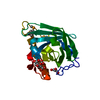
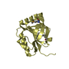
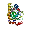
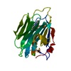
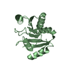
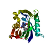
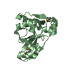
| Title | The M23 peptidase domain of the Staphylococcal phage 2638A endolysin | ||||||
 Components Components | ORF007 | ||||||
 Keywords Keywords | VIRAL PROTEIN / zinc metallopeptidase / bacteriophage / endolysin / M23 peptidase | ||||||
| Function / homology |  Function and homology information Function and homology informationsymbiont-mediated cytolysis of host cell / N-acetylmuramoyl-L-alanine amidase / N-acetylmuramoyl-L-alanine amidase activity / peptidoglycan catabolic process / metalloendopeptidase activity / defense response to bacterium / proteolysis / metal ion binding Similarity search - Function | ||||||
| Biological species |  Staphylococcus phage 2638A (virus) Staphylococcus phage 2638A (virus) | ||||||
| Method |  X-RAY DIFFRACTION / SYNCHROTRON / MOLECULAR REPLACEMENT / Resolution: 2.3 Å X-RAY DIFFRACTION / SYNCHROTRON / MOLECULAR REPLACEMENT / Resolution: 2.3 Å | ||||||
 Authors Authors | Dunne, M. / Ernst, P. / Sobieraj, A. / Pluckthun, A. / Loessner, M.J. | ||||||
 Citation Citation | Journal: To Be Published Title: (CASP target) Crystal structure of the M23 peptidase domain of Staphylococcal phage 2638A endolysin Authors: Sobieraj, A. / Dunne, M. / Ernst, P. / Pluckthun, A. / Loessner, M.J. | ||||||
| History |
|
- Structure visualization
| Structure viewer | Molecule: MolmilJmol/JSmol |
|---|
- Downloads & links
-Download
| PDBx/mmCIF format | 6yj1.cif.gz | 100.3 KB | Display | PDBx/mmCIF format |
|---|---|---|---|---|
| PDB format | pdb6yj1.ent.gz | 61.1 KB | Display | PDB format |
| PDBx/mmJSON format | 6yj1.json.gz | Tree view | PDBx/mmJSON format | |
| Others |  Other downloads Other downloads |
-Validation report
| Arichive directory | https://data.pdbj.org/pub/pdb/validation_reports/yj/6yj1ftp://data.pdbj.org/pub/pdb/validation_reports/yj/6yj1 | HTTPS FTP |
|---|
-Related structure data
| Related structure data |  4lxcS S: Starting model for refinement |
|---|---|
| Similar structure data |


-Links
 PDBj
PDBj- Assembly
| Deposited unit | 
| ||||||||||||||||||||||||
|---|---|---|---|---|---|---|---|---|---|---|---|---|---|---|---|---|---|---|---|---|---|---|---|---|---|
| 1 | 
| ||||||||||||||||||||||||
| 2 | 
| ||||||||||||||||||||||||
| Unit cell |
| ||||||||||||||||||||||||
| Noncrystallographic symmetry (NCS) | NCS domain:
NCS domain segments:
|
-Components
| #1: Protein | Mass: 21286.445 Da / Num. of mol.: 2 Source method: isolated from a genetically manipulated source Source: (gene. exp.) Staphylococcus phage 2638A (virus) / Production host:  #2: Chemical |   Mass: 65.409 Da / Num. of mol.: 2 / Source method: obtained synthetically / Formula: Zn Mass: 65.409 Da / Num. of mol.: 2 / Source method: obtained synthetically / Formula: Zn#3: Water | ChemComp-HOH / |  Mass: 18.015 Da / Num. of mol.: 120 / Source method: isolated from a natural source / Formula: H2O Mass: 18.015 Da / Num. of mol.: 120 / Source method: isolated from a natural source / Formula: H2OHas ligand of interest | N | Has protein modification | Y | |
|---|
-Experimental details
-Experiment
| Experiment | Method: X-RAY DIFFRACTION / Number of used crystals: 1 |
|---|
- Sample preparation
| Crystal | Density Matthews: 2.48 Å3/Da / Density % sol: 50.33 % |
|---|---|
| Crystal grow | Temperature: 292 K / Method: vapor diffusion, hanging drop / pH: 7.4 Details: Protein buffer: 20 mM Tris, 150 mM NaCl, pH 7.4 Crystallization buffer: 0.2 M sodium chloride, 30 % (v/v) PEG 300, pH 5.7 |
-Data collection
| Diffraction | Mean temperature: 100 K / Serial crystal experiment: N |
|---|---|
| Diffraction source | Source: SYNCHROTRON / Site: SLS  / Beamline: X06SA / Wavelength: 1 Å / Beamline: X06SA / Wavelength: 1 Å |
| Detector | Type: DECTRIS EIGER X 16M / Detector: PIXEL / Date: May 5, 2019 |
| Radiation | Protocol: SINGLE WAVELENGTH / Monochromatic (M) / Laue (L): M / Scattering type: x-ray |
| Radiation wavelength | Wavelength: 1 Å / Relative weight: 1 |
| Reflection | Resolution: 2.3→39.43 Å / Num. obs: 17866 / % possible obs: 95.06 % / Redundancy: 6.01 % / Biso Wilson estimate: 29.08 Å2 / CC1/2: 0.986 / CC star: 0.996 / Rmerge(I) obs: 0.1612 / Rpim(I) all: 0.0696 / Rrim(I) all: 0.1761 / Net I/σ(I): 7.59 |
| Reflection shell | Resolution: 2.3→2.382 Å / Redundancy: 5.74 % / Rmerge(I) obs: 0.5327 / Mean I/σ(I) obs: 4.1 / Num. unique obs: 1664 / CC1/2: 0.911 / CC star: 0.976 / Rpim(I) all: 0.235 / Rrim(I) all: 0.585 / % possible all: 89.46 |
- Processing
| Software |
| |||||||||||||||||||||||||||||||||||||||||||||||||
|---|---|---|---|---|---|---|---|---|---|---|---|---|---|---|---|---|---|---|---|---|---|---|---|---|---|---|---|---|---|---|---|---|---|---|---|---|---|---|---|---|---|---|---|---|---|---|---|---|---|---|
| Refinement | Method to determine structure: MOLECULAR REPLACEMENT Starting model: 4LXC Resolution: 2.3→39.43 Å / SU ML: 0.2541 / Cross valid method: FREE R-VALUE / σ(F): 1.36 / Phase error: 40.2503
| |||||||||||||||||||||||||||||||||||||||||||||||||
| Solvent computation | Shrinkage radii: 0.9 Å / VDW probe radii: 1.11 Å | |||||||||||||||||||||||||||||||||||||||||||||||||
| Displacement parameters | Biso mean: 32 Å2 | |||||||||||||||||||||||||||||||||||||||||||||||||
| Refinement step | Cycle: LAST / Resolution: 2.3→39.43 Å
| |||||||||||||||||||||||||||||||||||||||||||||||||
| Refine LS restraints |
| |||||||||||||||||||||||||||||||||||||||||||||||||
| LS refinement shell |
|
