 Movie
Movie Controller

[English] 日本語
 Yorodumi
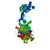
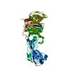
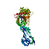
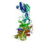
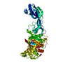
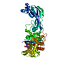
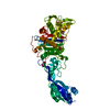
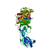
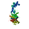
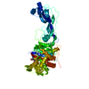
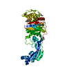
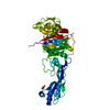
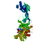
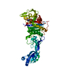
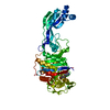
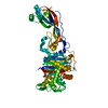
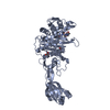
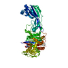
- PDB-7au1: Structure of P. aeruginosa PBP3 in complex with a benzoxaborole (... -
+ Open data

- Basic information
| Entry | Database: PDB / ID: 7au1 | |||||||||
|---|---|---|---|---|---|---|---|---|---|---|
| Title | Structure of P. aeruginosa PBP3 in complex with a benzoxaborole (Compound 12) | |||||||||
 Components Components | Peptidoglycan D,D-transpeptidase FtsI | |||||||||
 Keywords Keywords | HYDROLASE / D / D-transpeptidase / boron-binding / divalency | |||||||||
| Function / homology |  Function and homology information Function and homology informationpeptidoglycan glycosyltransferase activity / serine-type D-Ala-D-Ala carboxypeptidase / serine-type D-Ala-D-Ala carboxypeptidase activity / division septum assembly / FtsZ-dependent cytokinesis / penicillin binding / peptidoglycan biosynthetic process / cell wall organization / regulation of cell shape / proteolysis / plasma membrane Similarity search - Function | |||||||||
| Biological species |   Pseudomonas aeruginosa (bacteria) Pseudomonas aeruginosa (bacteria) | |||||||||
| Method |  X-RAY DIFFRACTION / SYNCHROTRON / MOLECULAR REPLACEMENT / molecular replacement / Resolution: 1.36 Å X-RAY DIFFRACTION / SYNCHROTRON / MOLECULAR REPLACEMENT / molecular replacement / Resolution: 1.36 Å | |||||||||
 Authors Authors | Newman, H. / Bellini, B. / Dowson, C.G. | |||||||||
| Funding support |  United Kingdom, 1items United Kingdom, 1items
| |||||||||
 Citation Citation | Journal: J.Med.Chem. / Year: 2021 Title: High-Throughput Crystallography Reveals Boron-Containing Inhibitors of a Penicillin-Binding Protein with Di- and Tricovalent Binding Modes. Authors: Newman, H. / Krajnc, A. / Bellini, D. / Eyermann, C.J. / Boyle, G.A. / Paterson, N.G. / McAuley, K.E. / Lesniak, R. / Gangar, M. / von Delft, F. / Brem, J. / Chibale, K. / Schofield, C.J. / Dowson, C.G. | |||||||||
| History |
|
- Structure visualization
| Structure viewer | Molecule: MolmilJmol/JSmol |
|---|
- Downloads & links
-Download
| PDBx/mmCIF format | 7au1.cif.gz | 223.8 KB | Display | PDBx/mmCIF format |
|---|---|---|---|---|
| PDB format | pdb7au1.ent.gz | 171.1 KB | Display | PDB format |
| PDBx/mmJSON format | 7au1.json.gz | Tree view | PDBx/mmJSON format | |
| Others |  Other downloads Other downloads |
-Validation report
| Arichive directory | https://data.pdbj.org/pub/pdb/validation_reports/au/7au1ftp://data.pdbj.org/pub/pdb/validation_reports/au/7au1 | HTTPS FTP |
|---|
-Related structure data
| Related structure data | 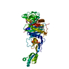 7atmC  7atoC  7atwC 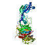 7atxC 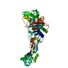 7au0C 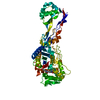 7au8C 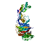 7au9C 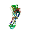 7aubC 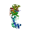 7auhC 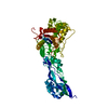 6hzrS C: citing same article ( S: Starting model for refinement |
|---|---|
| Similar structure data |


-Links
 PDBj
PDBj
- Assembly
| Deposited unit | 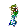
| ||||||||
|---|---|---|---|---|---|---|---|---|---|
| 1 |
| ||||||||
| Unit cell |
|
-Components
-Protein , 1 types, 1 molecules A
| #1: Protein | Mass: 58027.047 Da / Num. of mol.: 1 Source method: isolated from a genetically manipulated source Source: (gene. exp.) Pseudomonas aeruginosa (bacteria)Strain: ATCC 15692 / DSM 22644 / CIP 104116 / JCM 14847 / LMG 12228 / 1C / PRS 101 / PAO1 Gene: ftsI, pbpB, PA4418 / Plasmid: pET47b / Production host: References: UniProt: G3XD46, serine-type D-Ala-D-Ala carboxypeptidase |
|---|
-Non-polymers , 5 types, 411 molecules 


| #2: Chemical |  Mass: 78.133 Da / Num. of mol.: 2 / Source method: obtained synthetically / Formula: C2H6OS / Comment: DMSO, precipitant*YM Mass: 78.133 Da / Num. of mol.: 2 / Source method: obtained synthetically / Formula: C2H6OS / Comment: DMSO, precipitant*YM#3: Chemical |  Mass: 106.120 Da / Num. of mol.: 3 / Source method: obtained synthetically / Formula: C4H10O3 Mass: 106.120 Da / Num. of mol.: 3 / Source method: obtained synthetically / Formula: C4H10O3#4: Chemical | ChemComp-S08 / |  Mass: 368.148 Da / Num. of mol.: 1 / Source method: obtained synthetically / Formula: C18H17BN2O6 / Feature type: SUBJECT OF INVESTIGATION Mass: 368.148 Da / Num. of mol.: 1 / Source method: obtained synthetically / Formula: C18H17BN2O6 / Feature type: SUBJECT OF INVESTIGATION#5: Chemical | ChemComp-GOL /  Mass: 92.094 Da / Num. of mol.: 5 / Source method: obtained synthetically / Formula: C3H8O3 Mass: 92.094 Da / Num. of mol.: 5 / Source method: obtained synthetically / Formula: C3H8O3#6: Water | ChemComp-HOH / | Mass: 18.015 Da / Num. of mol.: 400 / Source method: isolated from a natural source / Formula: H2O |
|---|
-Details
| Has ligand of interest | Y |
|---|---|
| Has protein modification | Y |
-Experimental details
-Experiment
| Experiment | Method: X-RAY DIFFRACTION / Number of used crystals: 1 |
|---|
- Sample preparation
| Crystal | Density Matthews: 2.12 Å3/Da / Density % sol: 41.96 % |
|---|---|
| Crystal grow | Temperature: 293 K / Method: vapor diffusion, sitting drop / pH: 6 Details: 25% (w/v) polyethylene glycol 3,350, 0.1M Bis-Tris propane pH 6 and 1% (w/v) protamine sulphate |
-Data collection
| Diffraction | Mean temperature: 100 K / Serial crystal experiment: N |
|---|---|
| Diffraction source | Source: SYNCHROTRON / Site: Diamond / Beamline: I04-1 / Wavelength: 0.91587 Å |
| Detector | Type: DECTRIS PILATUS3 6M / Detector: PIXEL / Date: Apr 7, 2019 / Details: Mirrors |
| Radiation | Monochromator: M / Protocol: SINGLE WAVELENGTH / Monochromatic (M) / Laue (L): M / Scattering type: x-ray |
| Radiation wavelength | Wavelength: 0.91587 Å / Relative weight: 1 |
| Reflection | Resolution: 1.36→60.039 Å / Num. obs: 69775 / % possible obs: 91.7 % / Redundancy: 8.1 % / CC1/2: 0.999 / Rmerge(I) obs: 0.072 / Rpim(I) all: 0.026 / Rrim(I) all: 0.077 / Net I/σ(I): 15.1 |
| Reflection shell | Resolution: 1.36→1.491 Å / Redundancy: 5.7 % / Rmerge(I) obs: 0.85 / Num. unique obs: 3488 / Rpim(I) all: 0.381 / Rrim(I) all: 0.935 / % possible all: 62.5 |
-Phasing
| Phasing | Method: molecular replacement |
|---|
- Processing
| Software |
| |||||||||||||||||||||||||||||||||||||||||||||||||||||||||||||||||||||||||||||||||||||||||||||||||||||||||||||||||||||||||||||||||||||||||||||||||||||||||||||||||||||
|---|---|---|---|---|---|---|---|---|---|---|---|---|---|---|---|---|---|---|---|---|---|---|---|---|---|---|---|---|---|---|---|---|---|---|---|---|---|---|---|---|---|---|---|---|---|---|---|---|---|---|---|---|---|---|---|---|---|---|---|---|---|---|---|---|---|---|---|---|---|---|---|---|---|---|---|---|---|---|---|---|---|---|---|---|---|---|---|---|---|---|---|---|---|---|---|---|---|---|---|---|---|---|---|---|---|---|---|---|---|---|---|---|---|---|---|---|---|---|---|---|---|---|---|---|---|---|---|---|---|---|---|---|---|---|---|---|---|---|---|---|---|---|---|---|---|---|---|---|---|---|---|---|---|---|---|---|---|---|---|---|---|---|---|---|---|---|
| Refinement | Method to determine structure: MOLECULAR REPLACEMENT Starting model: 6HZR Resolution: 1.36→60.039 Å / Cor.coef. Fo:Fc: 0.973 / Cor.coef. Fo:Fc free: 0.951 / WRfactor Rfree: 0.199 / WRfactor Rwork: 0.143 / SU B: 3.811 / SU ML: 0.061 / Average fsc free: 0.9407 / Average fsc work: 0.9566 / Cross valid method: FREE R-VALUE / ESU R: 0.096 / ESU R Free: 0.084 Details: Hydrogens have been added in their riding positions
| |||||||||||||||||||||||||||||||||||||||||||||||||||||||||||||||||||||||||||||||||||||||||||||||||||||||||||||||||||||||||||||||||||||||||||||||||||||||||||||||||||||
| Solvent computation | Ion probe radii: 0.8 Å / Shrinkage radii: 0.8 Å / VDW probe radii: 1.2 Å / Solvent model: MASK BULK SOLVENT | |||||||||||||||||||||||||||||||||||||||||||||||||||||||||||||||||||||||||||||||||||||||||||||||||||||||||||||||||||||||||||||||||||||||||||||||||||||||||||||||||||||
| Displacement parameters | Biso mean: 20.581 Å2
| |||||||||||||||||||||||||||||||||||||||||||||||||||||||||||||||||||||||||||||||||||||||||||||||||||||||||||||||||||||||||||||||||||||||||||||||||||||||||||||||||||||
| Refinement step | Cycle: LAST / Resolution: 1.36→60.039 Å
| |||||||||||||||||||||||||||||||||||||||||||||||||||||||||||||||||||||||||||||||||||||||||||||||||||||||||||||||||||||||||||||||||||||||||||||||||||||||||||||||||||||
| Refine LS restraints |
| |||||||||||||||||||||||||||||||||||||||||||||||||||||||||||||||||||||||||||||||||||||||||||||||||||||||||||||||||||||||||||||||||||||||||||||||||||||||||||||||||||||
| LS refinement shell | Resolution: 1.36→1.395 Å
|
